. 2000 Nov 21;97(24):13051-6.
doi: 10.1073/pnas.220425797.

Essential role of Glu-C66 for menaquinol oxidation indicates transmembrane electrochemical potential generation by Wolinella succinogenes fumarate reductase

Affiliations

Essential role of Glu-C66 for menaquinol oxidation indicates transmembrane electrochemical potential generation by Wolinella succinogenes fumarate reductase

C R Lancaster et al. Proc Natl Acad Sci U S A. .

Abstract

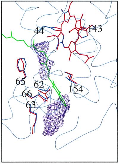
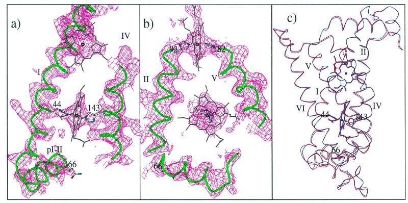
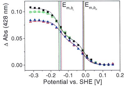
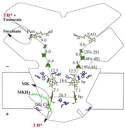
Quinol:fumarate reductase (QFR) is a membrane protein complex that couples the reduction of fumarate to succinate to the oxidation of quinol to quinone, in a reaction opposite to that catalyzed by the related enzyme succinate:quinone reductase (succinate dehydrogenase). In the previously determined structure of QFR from Wolinella succinogenes, the site of fumarate reduction in the flavoprotein subunit A of the enzyme was identified, but the site of menaquinol oxidation was not. In the crystal structure, the acidic residue Glu-66 of the membrane spanning, diheme-containing subunit C lines a cavity that could be occupied by the substrate menaquinol. Here we describe that, after replacement of Glu-C66 with Gln by site-directed mutagenesis, the resulting mutant is unable to grow on fumarate and the purified enzyme lacks quinol oxidation activity. X-ray crystal structure analysis of the Glu-C66-->Gln variant enzyme at 3.1-A resolution rules out any major structural changes compared with the wild-type enzyme. The oxidation-reduction potentials of the heme groups are not significantly affected. We conclude that Glu-C66 is an essential constituent of the menaquinol oxidation site. Because Glu-C66 is oriented toward a cavity leading to the periplasm, the release of two protons on menaquinol oxidation is expected to occur to the periplasm, whereas the uptake of two protons on fumarate reduction occurs from the cytoplasm. Thus our results indicate that the reaction catalyzed by W. succinogenes QFR generates a transmembrane electrochemical potential.


Figures

Figure 1
Figure 1
A working hypothesis. Two distal cavities (purple) in subunit C of the original structure of W. succinogenes QFR (PDB entry 1QLA) as detected with the program voidoo (33) and a working model of menaquinol binding (green) are shown. To accommodate the quinol head group in its current tentative position between the cavities, amino acid side-chain movements from their original positions (blue) to positions drawn in red are required as derived from energy minimization simulations with cns. The heme group shown is the distal heme bD. In this orientation, the periplasm is at the bottom and the rest of the QFR complex extends beyond the top and the right of the figure. Figs. 1, 2, and 4 were prepared with a version of molscript (34) modified for color ramping (35) and map drawing (36) capabilities.
Figure 2
Figure 2
The crystal structure of Glu-C66 → Gln QFR. (a and b) Representative sections of the 2|Fo|−|Fc| composite-omit electron density map, contoured at 1.0 standard deviations (σ) above the mean density of the map. Shown are the two heme groups, bP (Upper) and bD (Lower), in subunit C, as well as the periplasmic helix pI-II with its C-terminal extension containing the exchanged residue C66 (a and b). Drawn in green are the Cα traces of the transmembrane helices I and IV (a) and II and V (b) that form the four-helix bundle enclosing the two heme groups (5). To reduce overlap, the upper ends of helices IV, II, and V have been omitted. (c) Comparison of the heme positions and the Cα traces for the C subunits of wild-type QFR (blue, PDB entry 1QLA) and Glu-C66 → Gln QFR (red). The rms deviation for the 254 Cα atoms in the C subunit models is 0.44 Å.
Figure 3
Figure 3
The oxidation-reduction potentials of heme bH and bL in wild-type QFR and Glu-C66 → Gln QFR. Electrochemical redox titrations of wild-type QFR (squares) and Glu-C66 → Gln QFR (triangles). The amplitude of the Soret band maximum at 428 nm is plotted as a function of the applied potential: open symbols correspond to an oxidative titration, full symbols correspond to a reductive titration. To avoid overlap, absorbance differences are displayed as measured and have not been scaled according to the cytochrome b concentration. The black and green curves show a reversible titration of wild-type QFR, blue and red refer to Glu-C66 → Gln QFR. Fitting to calculated Nernst curves yielded the following individual values for the midpoint potentials Em of hemes bL and bH, with the first value determined from the reductive titration and the second from the oxidative titration. For wild-type QFR: Em,bL = −155 mV and −149 mV; Em,bH = −8 mV and −10 mV. For Glu-C66 → Gln QFR: Em,bL = −145 mV and −139 mV; Em,bH = −16 mV and −12 mV, as indicated by the vertical colored lines. SHE, standard hydrogen electrode.
Figure 4
Figure 4
Transmembrane electrochemical potential generation by W. succinogenes QFR coupling the two-electron oxidation of menaquinol (MKH2) to menaquinone (MK) to the two-electron reduction of fumarate to succinate. The positive (+) and negative (−) sides of the membrane are indicated. The prosthetic groups of the QFR dimer are displayed (coordinate set PDB entry 1QLA; ref. 5). Distances between prosthetic groups are edge-to-edge distances in Å as defined in ref. . Distances shorter than 14 Å (i.e., within one QFR monomer, but not between the two monomers of the dimer) are considered to be relevant for physiological electron transfer. Also drawn are the side chains of Glu-C66 (in red) and of the subunit C Trp residues (purple). The latter are markers for the hydrophobic surface-to-polar transition zone of the membrane. The position of bound fumarate (Fum) is taken from PDB entry 1QLB (5). The tentative model of menaquinol binding (drawn in green) is taken from Fig. 1. Its edge-to-edge distance to heme bD is 6.7 Å.

References

    1. Saraste M. Science. 1999;283:1488–1493. - PubMed
    1. Kröger A. Biochim Biophys Acta. 1978;505:129–145. - PubMed
    1. Kröger A, Geisler V, Lemma E, Theis F, Lenger R. Arch Microbiol. 1992;158:311–314.
    1. Hägerhäll C. Biochim Biophys Acta. 1997;1320:107–141. - PubMed
    1. Lancaster C R D, Kröger A, Auer M, Michel H. Nature (London) 1999;402:377–385. - PubMed

Publication types

Associated data

LinkOut - more resources