

RSK2 represses HSF1 activation during heat shock
- PMID: 11189448
- PMCID: PMC312873
- DOI: 10.1379/1466-1268(2000)005<0432:rrhadh>2.0.co;2
RSK2 represses HSF1 activation during heat shock
Abstract
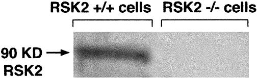
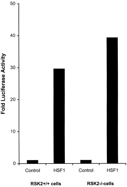
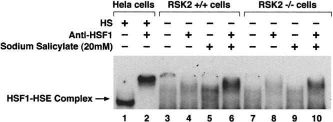
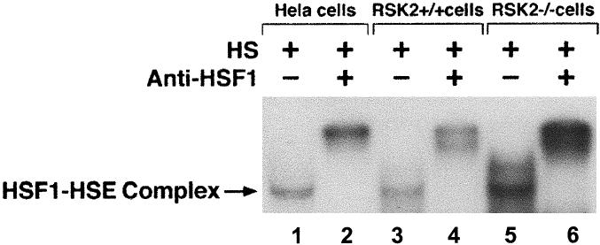
Heat shock transcription factor 1(HSF1) activation is a multistep process. The conversion of a latent cytoplasmic form to a nuclear, DNA binding state appears to be activated by nonsteroidal anti-inflammatory drugs. In previous studies, we showed that HSF 1 is phosphorylated by the protein kinase RSK2 in vitro and that this effect is inhibited by nonsteroidal anti-inflammatory drugs at the concentration that leads to the activation of HSF1 in vivo (Stevenson et al 1999). In the present study, using cells from a patient with Coffin-Lowry syndrome (deficient in RSK2), we demonstrate that RSK2 slightly represses activation of HSF1 in vivo at 37 degrees C. In Coffin-Lowry syndrome cells, HSF1-HSE DNA binding activity after treatment with sodium salicylate was slightly higher than that in untreated cells, indicating that although RSK2 is involved in HSF1 regulation, it is not the unique protein kinase that suppresses HSF1-HSE binding activity at 37 degrees C. However, heat shock treatment resulted in significantly higher HSF1-HSE binding activity in Coffin-Lowry syndrome cells as compared with normal controls, suggesting that RSK2 represses HSF1-HSE binding activity during heat shock.
Figures
Similar articles
-
Implication of reactive oxygen species, ERK1/2, and p38MAPK in sodium salicylate-induced heat shock protein 72 expression in C6 glioma cells.Int J Mol Med. 2005 Nov;16(5):841-9. Int J Mol Med. 2005. PMID: 16211253
-
Salicylate triggers heat shock factor differently than heat.J Biol Chem. 1995 Oct 13;270(41):24489-95. doi: 10.1074/jbc.270.41.24489. J Biol Chem. 1995. PMID: 7592665
-
Activation of heat shock factor 1 DNA binding precedes stress-induced serine phosphorylation. Evidence for a multistep pathway of regulation.J Biol Chem. 1996 Feb 16;271(7):3355-8. doi: 10.1074/jbc.271.7.3355. J Biol Chem. 1996. PMID: 8631933
-
Heat shock factor 1 (HSF1)-targeted anticancer therapeutics: overview of current preclinical progress.Expert Opin Ther Targets. 2019 May;23(5):369-377. doi: 10.1080/14728222.2019.1602119. Epub 2019 Apr 7. Expert Opin Ther Targets. 2019. PMID: 30931649 Review.
-
New insights into the mechanism of heat shock response activation.Cell Mol Life Sci. 2008 Mar;65(6):855-61. doi: 10.1007/s00018-008-7458-y. Cell Mol Life Sci. 2008. PMID: 18239856 Free PMC article. Review.
Cited by
-
Role of Heat Shock Factors in Stress-Induced Transcription: An Update.Methods Mol Biol. 2023;2693:25-38. doi: 10.1007/978-1-0716-3342-7_3. Methods Mol Biol. 2023. PMID: 37540424
-
Role of Heat Shock Factors in Stress-Induced Transcription.Methods Mol Biol. 2018;1709:23-34. doi: 10.1007/978-1-4939-7477-1_2. Methods Mol Biol. 2018. PMID: 29177648 Free PMC article.
-
Signal Transduction Pathways Leading to Heat Shock Transcription.Sign Transduct Insights. 2010;2:13-24. doi: 10.4137/STI.S3994. Sign Transduct Insights. 2010. PMID: 21687820 Free PMC article.
-
A transcription cofactor required for the heat-shock response.EMBO Rep. 2008 Jul;9(7):662-9. doi: 10.1038/embor.2008.70. Epub 2008 May 2. EMBO Rep. 2008. PMID: 18451878 Free PMC article.
-
Regulation of molecular chaperone gene transcription involves the serine phosphorylation, 14-3-3 epsilon binding, and cytoplasmic sequestration of heat shock factor 1.Mol Cell Biol. 2003 Sep;23(17):6013-26. doi: 10.1128/MCB.23.17.6013-6026.2003. Mol Cell Biol. 2003. PMID: 12917326 Free PMC article.
References
-
- Biancalana V, Trivier E, Weber C, et al. Construction of a high-resolution linkage map for Xp22.1-p22.2 and refinement of the genetic localization of the Coffin-Lowry syndrome gene. Genomics. 1994;22:617–625. - PubMed
-
- Bird H, Collins AL, Oley C, Lindsay S. Crossover analysis in a British family suggests that Coffin-Lowry syndrome maps to a 3.4-cM interval in Xp22. Am J Med Genet. 1995;59:512–516. - PubMed
-
- Cahill CM, Waterman WR, Xie Y, Auron PE, Calderwood SK. Transcriptional repression of the prointerleukin 1beta gene by heat shock factor 1. J Biol Chem. 1996;271:24874–24879. - PubMed
-
- Calderwood SK 1995 Molecular strategies for sensing and responding to stress. Proceedings of the 86th Annual Meeting of the American Association for Cancer Research. 65682.
Publication types
MeSH terms
Substances
Grants and funding
LinkOut - more resources
Full Text Sources