

Disruption of cell cycle kinetics by benzo[a]pyrene: inverse expression patterns of BRCA-1 and p53 in MCF-7 cells arrested in S and G2
- PMID: 11191113
- PMCID: PMC1507983
- DOI: 10.1038/sj.neo.7900104
Disruption of cell cycle kinetics by benzo[a]pyrene: inverse expression patterns of BRCA-1 and p53 in MCF-7 cells arrested in S and G2
Abstract
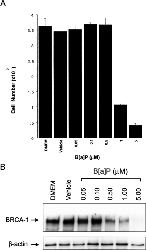
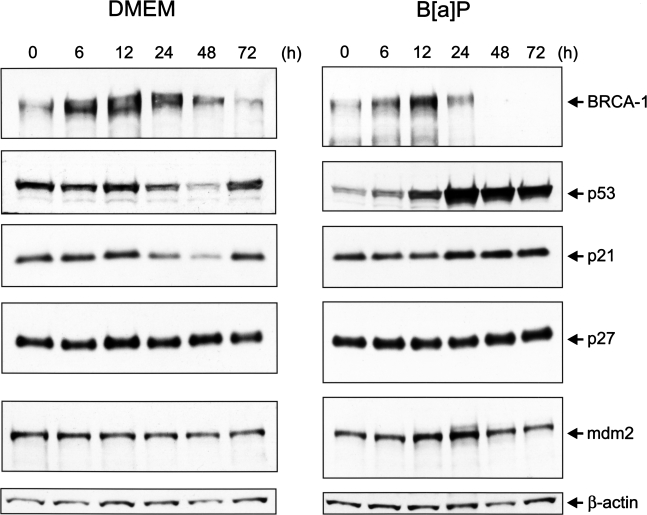
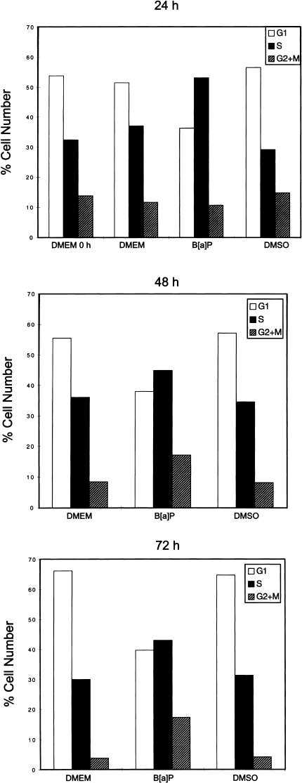
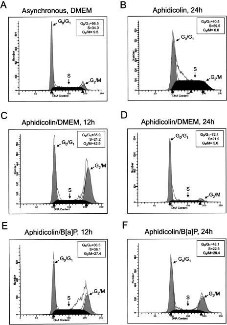
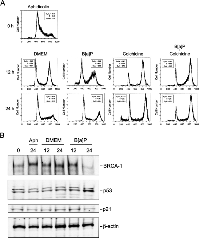
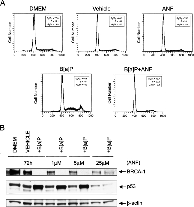
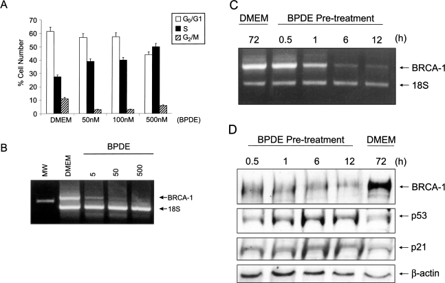
The effects of a ligand of the aromatic hydrocarbon receptor (AhR), benzo[a] pyrene (B[ a]P), and its metabolite, BPDE (7r,8t-dihydroxy-9t,10t-epoxy-7,8,9,10-tetrahydro-benzo[a]pyrene), on BRCA-1 levels and cell cycle kinetics were determined in MCF-7 breast cancer cells. Exposure of asynchronous MCF-7 cells for 72 hours to a non-cytotoxic dose of 0.5 microM B[a]P triggered a three-fold reduction in BRCA-1 protein. In MCF-7 cells resistant (20% to 30%) to genotoxic concentrations of B[a]P (1 to 5 microM), the loss of BRCA-1 protein was coupled with pausing in S-phase and G2/M, and accumulation of p53, mdm2 and p21. Treatment of MCF-7 cells synchronized in S-phase (72%) with B[a]P prolonged the arrest in S-phase, although this checkpoint was transient since cells resumed to G2/M after 12 hours with reduced levels of BRCA-1. In these cells, levels of p53 were increased, whereas the cellular content of p21 remained unaltered. In contrast, the co-treatment with the AhR antagonist, alpha-naphthoflavone (ANF), abrogated the deleterious effects of B[a]P on BRCA-1 expression, while preventing the accumulation of p53 and disruption of cell cycle profile. These findings suggest that the AhR mediated the inverse expression patterns of BRCA-1 and p53 upon exposure to B[a]P. The treatment with BPDE induced S-phase arrest and reduced BRCA-1 mRNA levels. The negative effects of BPDE on BRCA-1 expression were not transient since removal of BPDE did not allow complete reversal of the repression. These cumulative data suggest that the B[a]P metabolite, BPDE, may play a key role in disruption of BRCA-1 expression and cell cycle kinetics in breast epithelial cells.
Figures
Similar articles
-
Activation of the aromatic hydrocarbon receptor pathway is not sufficient for transcriptional repression of BRCA-1: requirements for metabolism of benzo[a]pyrene to 7r,8t-dihydroxy-9t,10-epoxy-7,8,9,10-tetrahydrobenzo[a]pyrene.Cancer Res. 2002 Jan 1;62(1):113-21. Cancer Res. 2002. PMID: 11782367
-
Inhibition of BRCA-1 expression by benzo[a]pyrene and its diol epoxide.Mol Carcinog. 1999 Oct;26(2):100-18. doi: 10.1002/(sici)1098-2744(199910)26:2<100::aid-mc5>3.0.co;2-1. Mol Carcinog. 1999. PMID: 10506754
-
Epigenetics of breast cancer: polycyclic aromatic hydrocarbons as risk factors.Environ Mol Mutagen. 2002;39(2-3):235-44. doi: 10.1002/em.10051. Environ Mol Mutagen. 2002. PMID: 11921194
-
A benzo[a]pyrene-induced cell cycle checkpoint resulting in p53-independent G1 arrest in 3T3 fibroblasts.J Biol Chem. 1997 Jan 31;272(5):2762-9. doi: 10.1074/jbc.272.5.2762. J Biol Chem. 1997. PMID: 9006915
-
The ligand status of the aromatic hydrocarbon receptor modulates transcriptional activation of BRCA-1 promoter by estrogen.Cancer Res. 2006 Feb 15;66(4):2224-32. doi: 10.1158/0008-5472.CAN-05-1619. Cancer Res. 2006. PMID: 16489025
Cited by
-
STIM2 is involved in the regulation of apoptosis and the cell cycle in normal and malignant monocytic cells.Mol Oncol. 2024 Jun;18(6):1571-1592. doi: 10.1002/1878-0261.13584. Epub 2024 Jan 17. Mol Oncol. 2024. PMID: 38234211 Free PMC article.
-
The intervention mechanism of folic acid for benzo(a)pyrene toxic effects in vitro and in vivo.Eur J Cancer Prev. 2019 Jul;28(4):355-364. doi: 10.1097/CEJ.0000000000000461. Eur J Cancer Prev. 2019. PMID: 30020114 Free PMC article.
-
An estrogen receptor-alpha/p300 complex activates the BRCA-1 promoter at an AP-1 site that binds Jun/Fos transcription factors: repressive effects of p53 on BRCA-1 transcription.Neoplasia. 2005 Sep;7(9):873-82. doi: 10.1593/neo.05256. Neoplasia. 2005. PMID: 16229810 Free PMC article.
-
Time- and concentration-dependent changes in gene expression induced by benzo(a)pyrene in two human cell lines, MCF-7 and HepG2.BMC Genomics. 2006 Oct 16;7:260. doi: 10.1186/1471-2164-7-260. BMC Genomics. 2006. PMID: 17042939 Free PMC article.
-
Lack of association between GSTT1 polymorphism and endogenous or benzo[a]pyrene-induced sister chromatid exchanges as analyzed in metaphase or G2-phase lymphocytes.Mol Biol Rep. 2011 Aug;38(6):3959-66. doi: 10.1007/s11033-010-0513-4. Epub 2010 Nov 25. Mol Biol Rep. 2011. PMID: 21107716
References
-
- Gowen LC, Avrutskaya AV, Latour AM, Koller BH, Leadon SA. BRCA-1 required for transcription-coupled repair of oxidative damage. Science. 1998;286:804–810. - PubMed
-
- Scully R, Chen J, Plug A, Xiao Y, Waver D, Feunteun J, Ashley T, Livingston DM. Association of BRCA-1 with Rad51 in mitotic and meiotic cells. Cell. 1997;88:265–275. - PubMed
-
- Zhang H, Tombline G, Weber BL. BRCA-1, BRCA-2 and DNA damage response: collision or collusion. Cell. 1998;92:433–436. - PubMed
-
- Zhong Q, Chen C-F, Li S, Chen Y, Wang C-C, Xiao J, Chen P-L, Sharp ZD, Lee W-H. Association of BRCA-1 with the hRad50-hMre11-p95 complex and the DNA damage response. Science. 1999;285:747–750. - PubMed
-
- Hakem R, de la Pompa JL, Elia A, Potter J, Mak TW. Partial rescue of BRCA-1, (5–6) early embryonic lethality by p53 or p21 null mutation. Nat Genet. 1997;16:298–302. - PubMed
Publication types
MeSH terms
Substances
Grants and funding
LinkOut - more resources
Full Text Sources
Medical
Research Materials
Miscellaneous