. 2001 Mar 1;166(5):3266-76.
doi: 10.4049/jimmunol.166.5.3266.

MHC class I-related neonatal Fc receptor for IgG is functionally expressed in monocytes, intestinal macrophages, and dendritic cells

Affiliations

MHC class I-related neonatal Fc receptor for IgG is functionally expressed in monocytes, intestinal macrophages, and dendritic cells

X Zhu et al. J Immunol. .

Abstract

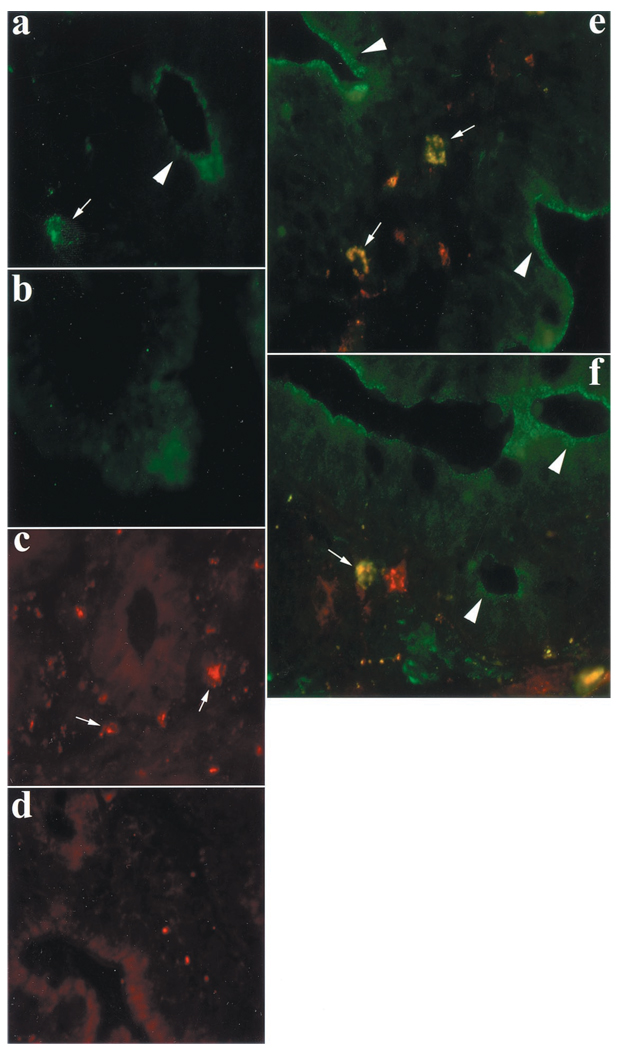
The neonatal Fc receptor (FcRn) for IgG, an MHC class I-related molecule, functions to transport IgG across polarized epithelial cells and protect IgG from degradation. However, little is known about whether FcRn is functionally expressed in immune cells. We show here that FcRn mRNA was identifiable in human monocytes, macrophages, and dendritic cells. FcRn heavy chain was detectable as a 45-kDa protein in monocytic U937 and THP-1 cells and in purified human intestinal macrophages, peripheral blood monocytes, and dendritic cells by Western blot analysis. FcRn colocalized in vivo with macrosialin (CD68) and Ncl-Macro, two macrophage markers, in the lamina propria of human small intestine. The heavy chain of FcRn was associated with the beta(2)-microglobulin (beta(2)m) light chain in U937 and THP-1 cells. FcRn bound human IgG at pH 6.0, but not at pH 7.5. This binding could be inhibited by human IgG Fc, but not Fab. FcRn could be detected on the cell surface of activated, but not resting, THP-1 cells. Furthermore, FcRn was uniformly present intracellularly in all blood monocytes and intestinal macrophages. FcRn was detectable on the cell surface of a significant fraction of monocytes at lower levels and on a small subset of tissue macrophages that expressed high levels of FcRn on the cell surface. These data show that FcRn is functionally expressed and its cellular distribution is regulated in monocytes, macrophages, and dendritic cells, suggesting that it may confer novel IgG binding functions upon these cell types relative to typical Fc gamma Rs: Fc gamma RI, Fc gamma RII, and Fc gamma RIII.


Figures

FIGURE 1
FIGURE 1
Detection of human FcRn heavy chain by immunoblotting with the mouse anti-α2-specific serum. Total cellular proteins (60 µg) from either HeLamock or HeLaFcRn + β2m transfectant were resolved on a 12% SDS-PAGE gel under reducing conditions. Immunoblotting was performed with the mouse anti-α2-specific serum and HRP-conjugated goat anti-mouse IgG, with detection accomplished by ECL. The arrow indicates the heavy chain of human FcRn. The Mr markers in kilodaltons are indicated on the right.
FIGURE 2
FIGURE 2
Purity of isolated dendritic cells and intestinal lamina propria macrophages. A, Primary lamina propria macrophages were isolated and purified from normal human jejunum as described in Materials and Methods and then analyzed by flow cytometry for the indicated surface Ags. Gates were set to include the total cell population. Insets display FACS profiles using the CD-specific Abs with the following purified control cells: CD103, intestinal lymphocytes; CD14, blood monocytes; CD3, blood lymphocytes; CD20, blood lymphocytes; and CD83, blood monocyte-derived dendritic cells. The FACS profiles are representative of lamina propria macrophages from intestinal tissue from a single donor (n = 6). The MFI is shown on the x-axis, and the relative cell number on the y-axis. B, Monocytes were isolated from PBMCs and stimulated with recombinant human GM-CSF and recombinant human IL-4 for 8 days. After 8 days, cells displaying dendritic morphology, as shown by forward and side scatter, and predominantly expressed CD1a and HLA-DR but which had lost most of the expression of the monocyte marker CD14 were isolated. Staining is shown with specific mAbs directly conjugated to PE.
FIGURE 3
FIGURE 3
RT-PCR amplification of FcRn cDNA from immune cells. First-strand cDNA was prepared as described in Materials and Methods. Amplified PCR products (800 bp) were electrophoresed in 1.2% agarose gels and stained with ethidium bromide. Similar PCR products amplified with a GAPDH-specific primer pair was also fractionated in 1.2% agarose gels as internal controls. The arrow indicates the location of the amplification products for human FcRn heavy chain (hFcRn) and GAPDH. The m.w. markers in base pairs are indicated on the left. SI, small intestine; Mϕ, macrophages; DC, dendritic cells.
FIGURE 4
FIGURE 4
FcRn expression in freshly isolated cells and cell lines. A, Detection of human FcRn protein in freshly isolated monocytes, small intestinal macrophages, dendritic cells, and cell lines by Western blot. SDS-PAGE gels were loaded with 60 µg of total protein per lane from the indicated sources, and the proteins were resolved under reducing conditions. The proteins were transferred onto nitrocellulose and probed with a rabbit anti-α2-specific serum and HRP-conjugated donkey anti-rabbit IgG used for development. The protein bands were visualized by ECL. The arrow indicates the location of the human FcRn heavy chain (hFcRn). B, Immunofluorescence staining of monocyte-like cell lines THP-1 and U937. The THP-1 and U937 cell lines were grown on glass coverslips, fixed with 3.7% paraformaldehyde, and permeabilized in 0.1% digitonin. Subsequently, the cells were incubated with mouse anti-α2-specific serum, followed by staining with a FITC-conjugated F(ab)2 goat anti-mouse Ab (panels C and E). HeLaFcRn + β2m cells were stained as positive controls (A). The U937 cell line was stained with normal mouse serum as negative control (panel G). The nucleus was stained with 4′6′-diamidino-2-phenylindole (panels B, D, F, and H) and photographed through a fluorescence microscope. Positive samples and negative controls were viewed using the same contrast and brightness settings. C, Detection of FcRn association with β2m in monocyte-like cell lines. Metabolically labeled THP-1 and U937 cells were immunoprecipitated with either rabbit anti-α2-specific serum or nonimmune serum and analyzed by SDS-PAGE and autoradiography. The 45- and 12-kDa bands were coprecipitated in the presence of hFcRn-specific immune serum, but not in the presence of nonimmune serum. The Mr markers in kilodaltons are indicted on the left. The locations of human FcRn heavy chain (hFcRn) and β2m are indicated by arrows.
FIGURE 5
FIGURE 5
Immunolocalization of FcRn in macrophages of the lamina propria in adult human small intestine. Frozen sections of tissue samples obtained from normal human jejunum were stained with either rabbit anti-FcRn Ab or anti-Ncl-Macro mAb. a, e, and f, arrowheads, Crypt and villus enterocytes show a punctuate staining pattern of FcRn expression visible at the apical plasma membrane and in the apical cytoplasm. a, arrow, A nearby resident lamina propria macrophage expresses FcRn. b, FcRn staining was not observed in the presence of an irrelevant antiserum. c, arrows, Abs against Ncl-Macro-stained lamina propria macrophages. d, Macrophage staining was absent in the presence of an irrelevant isotype-matched mAb. Double labeling with both anti-FcRn and anti-Ncl-Macro Abs revealed colocalization (yellow, arrow) of FcRn and Ncl-Macro in lamina propria macrophages of the villus (e) and crypt (f).
FIGURE 6
FIGURE 6
Detection of pH-dependent FcRn binding of IgG in macrophages and dendritic cells. IgG binding assays were performed at both pH 6.0 and 7.5 as described in Materials and Methods. The U937, THP-1, monocyte-derived dendritic cells, and intestinal macrophages were lysed in sodium phosphate buffer (pH 6.0 or 7.5) with 0.5% CHAPS. Approximately 0.5–1 mg of soluble proteins were incubated with human IgG-Sepharose at 4°C. The eluted proteins were subjected to 12% SDS-PAGE analysis under reducing conditions. Proteins were probed with a rabbit anti-α2-specific serum and developed with HRP-conjugated donkey anti-rabbit Abs with visualization by ECL. Lysates of HeLaFcRn + β2m were probed similarly as a positive control. The Mr markers in kilodaltons are indicated on the right. The location of the human FcRn heavy chain is indicated by an arrow.
FIGURE 7
FIGURE 7
Blockade of FcRn-mediated IgG binding by IgG Fc fragment. IgG binding assays were performed as described in Fig. 6. For blocking, 250–500 µg of human Fc or F(ab)2 were added to IgG-Sepharose beads before adding lysates from the THP-1 cell line. The eluted proteins were analyzed by 12% SDS-PAGE under reducing conditions, probed with a rabbit anti-α2-specific serum, and developed with HRP-conjugated secondary Abs with visualization by ECL. Lysates of HeLaFcRn + β2m were probed similarly as a positive control. The Mr markers in kilodaltons are indicated on the right.
FIGURE 8
FIGURE 8
Cellular distribution of FcRn expression patterns of FcRn on THP-1, monocytes, and macrophages. A, Surface biotinylation of FcRn on resting THP-1 and PMA-activated THP-1 cell lines. THP-1 cells were treated with 100 nm/ml PMA for 48 h. Cell surface proteins were biotinylated and solubilized at pH 6.0 as described in the text. Lysates were incubated with IgG-Sepharose beads. The eluted proteins were analyzed by SDS-PAGE electrophoresis under reducing conditions, blotted with streptavidin-HRP, and developed with ECL. This experiment was conducted in a duplicate sample with identical results. The specificity of the band identified in the avidin blot was provided by immunoprecipitation with an FcRn-specific Ab followed by avidin blotting (data not shown). B, Analysis of the surface and intracellular expression of FcRn on resting and PMA-activated THP-1 cells. Indirect immunofluorescence staining was performed on untreated cells or cells treated with PMA for the indicated time periods (hours). Cell surface and intracellular expression of FcRn on resting or activated THP-1 cells was described in Materials and Methods. Results are expressed as histograms of MFI (log scale) on the x-axis. The open peak represents staining of cells with the anti-hFcRn Ab, and the filled peak represents cells stained with irrelevant IgG. C, Expression of FcRn in monocytes and macrophages analyzed by flow cytometry. Cell surface and intracellular expression patterns of FcRn in either fixed or permeabilized blood monocytes and small intestinal macrophages were measured by flow cytometry. Cells were stained as described in Materials and Methods. Results are expressed as histograms of fluorescence intensity (log scale). The filled histograms represent staining of cells with anti-α2-specific serum, and the open histograms represent cells stained with irrelevant IgG. Values in the top right of each rectangle correspond to the proportion of cells stained with the anti-hFcRn Ab relative to the control Ab. The staining for macrophages and monocytes was conducted three times with similar results.

Similar articles

Cited by

References

    1. Simister NE, Rees AR. Isolation and characterization of an Fc receptor from neonatal rat small intestine. Eur. J. Immunol. 1985;15:733. - PubMed
    1. Simister NE, Mostov KE. An Fc receptor structurally related to MHC class I antigens. Nature. 1989;337:184. - PubMed
    1. Burmelster WP, Gastinel LN, Simister NE, Blum ML, Bjorkman PJ. Crystal structure at 2.2 Å resolution of the MHC-related neonatal Fc receptor. Nature. 1994;372:336. - PubMed
    1. Jones EA, Waldmann TA. The mechanism of intestinal uptake and transcellular transport of IgG in the neonatal rat. J. Clin. Invest. 1972;51:2916. - PMC - PubMed
    1. Abrahamson D, Rodewald R. Evidence for sorting of endocytic vesicle contents during the receptor-mediated transport of IgG across the newborn rat intestine. J. Cell Biol. 1981;91:270. - PMC - PubMed

Publication types

MeSH terms