. 2001 Feb;123(2):226-32.
doi: 10.1046/j.1365-2249.2001.01450.x.

Epitope focus, clonal composition and Th1 phenotype of the human CD4 response to the secretory mycobacterial antigen Ag85

Affiliations

Epitope focus, clonal composition and Th1 phenotype of the human CD4 response to the secretory mycobacterial antigen Ag85

M T Valle et al. Clin Exp Immunol. 2001 Feb.

Abstract

Lymphoproliferation of healthy donors was tested against mycobacterial antigens (PPD, Ag85, Ag85 peptides). All PPD responders recognized the secretory antigen Ag85 and the peptide specificity for Ag85B was defined. Peptide 91-108 was recognized by 85% of donors. In addition, all CD4 T cell lines generated from 12 donors against PPD or Ag85 responded to 91-108. When this peptide was used to generate T cell lines, the cells responded also to tuberculins from atypical mycobacterial species. Thus the cross-reactive peptide behaved as quasi-universal. The analysis of TCR-BV gene usage by cell lines showed that most Ag85-specific T cells correspond to 91-108-specific clonotypes. Intracytoplasmic staining of cell lines after phorbol myristate acetate stimulation resulted in dominance of interferon-gamma (IFN-gamma)-IL-4 double-positive cells, whereas antigen stimulation resulted in production of IFN-gamma only. The data show that peptide 91-108 is the major focus of the CD4 response to mycobacterial antigens in peripheral blood mononuclear cells and in T cell lines from PPD responders.


Figures

Fig. 1
Fig. 1
Left panel: response of 14 normal donors to Ag85B peptides. Responses with stimulation index (SI) >2 were scored as positive. Eleven individuals responded to PEP11. Right panel: response of 32 normal donors to PPD (□) and PEP11 (▪). Results are shown as SI (ratio between ct/min of antigen-stimulated wells and ct/min of control wells).
Fig. 2
Fig. 2
Peptide specificity of T cell lines. Left panel: 12 T cell lines generated by stimulation with PPD were tested with Ag85B peptides; 11/12 exhibited a stimulation index (SI) >2 with PEP11. Right panel: 14 T cell lines generated by stimulation with Ag85 were tested with the panel of peptides; 14/14 exhibited an SI >2 with PEP11.
Fig. 3
Fig. 3
Response to deleted peptides. Amino and carboxy deleted peptides derived from the sequence of PEP11 were tested on peripheral blood mononuclear cells (PBMC) (upper left panel), PPD-specific (upper right panel), Ag85-generated (lower left panel) and PEP11-generated T cell lines (lower right panel). The shortest peptide that retained the stimulatory ability of PEP11 with all of the T cells shown in the four panels was QTYKWETLLTSE (aa 93–104).
Fig. 4
Fig. 4
Recognition of PEP11 across different mycobacterial species. A T cell line induced by in vitro stimulation with PEP11 was tested with a panel of tuberculin preparations obtained from different species. The 93–104 sequence in the different species is shown with the corresponding substitutions. Proliferative responses comparable to those obtained with PEP11 and Ag85 were seen with all mycobacterial species tested here.
Fig. 5
Fig. 5
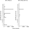
TCR-BV gene usage by PPD-specific lines. A PPD-specific line split and restimulated with PPD or with PEP11 for three cycles. The analysis of TCR-BV gene family usage showed that BV 18 was well represented, but flanked by other BV genes, in the PPD-restimulated line. In contrast, BV 18 was the dominant family in the PEP11-selected T cell line. Results are shown as pixel intensity of the ethidium bromide-stained bands of polymerase chain reaction products run on an agarose gel.
Fig. 6
Fig. 6
Intracytoplasmic staining of antigen-specific CD4 T cells. PPD-generated (left panel) and PEP11-generated (right panel) T cell lines were stimulated with PPD or PEP11 (or with phorbol myristate acetate (PMA) as a positive control) and stained for intracytoplasmic IFN-γ synthesis. Results are shown as percentage of positively stained T cells over total CD4 lymphocytes.

Similar articles

Cited by

References

    1. Fine PEM. The BCG story: lessons from the past and implications for the future. Rev Infect Dis. 1989;11:S353–9. - PubMed
    1. Young DB, Kaufmann SHE, Hermans PWM, Thole JER. Mycobacterial protein antigens: a compilation. Molec Microbiol. 1992;6:133–45. - PubMed
    1. Lamb JR, Lathigra R, Rothbard JB, Sweetser D, Young RA, Ivanyi J, Young DB. Identification of mycobacterial antigens recognized by T lymphocytes. Rev Infect Dis. 1989;11:S443–7. - PubMed
    1. Horwitz MA. A new TB vaccine. Immunologist. 1997;5:15–20.
    1. Kauffmann S. Immunity to intracellular bacteria. Annu Rev Immunol. 1993;11:129–63. - PubMed

Publication types

MeSH terms