. 2001 Feb;183(3):951-8.
doi: 10.1128/JB.183.3.951-958.2001.

Periplasmic transit and disulfide bond formation of the autotransported Shigella protein IcsA

Affiliations

Periplasmic transit and disulfide bond formation of the autotransported Shigella protein IcsA

L D Brandon et al. J Bacteriol. 2001 Feb.

Abstract

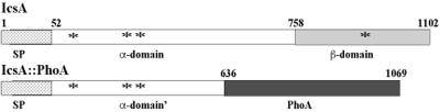
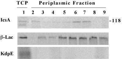
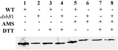
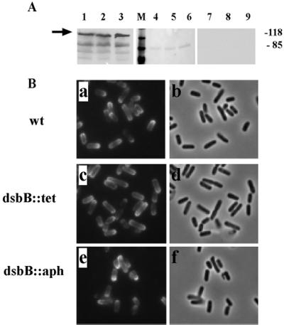
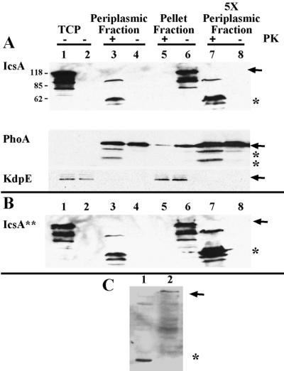
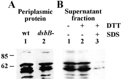
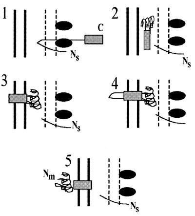
The Shigella outer membrane protein IcsA belongs to the family of type V secreted (autotransported) virulence factors. Members of this family mediate their own translocation across the bacterial outer membrane: the carboxy-terminal beta domain forms a beta barrel channel in the outer membrane through which the amino-terminal alpha domain passes. IcsA, which is localized at one pole of the bacterium, mediates actin assembly by Shigella, which is essential for bacterial intracellular movement and intercellular dissemination. Here, we characterize the transit of IcsA across the periplasm during its secretion. We show that an insertion in the dsbB gene, whose gene product mediates disulfide bond formation of many periplasmic intermediates, does not affect the surface expression or unipolar targeting of IcsA. However, IcsA forms one disulfide bond in the periplasm in a DsbA/DsbB-dependent fashion. Furthermore, cellular fractionation studies reveal that IcsA has a transient soluble periplasmic intermediate. Our data also suggest that IcsA is folded in a proteinase K-resistant state in the periplasm. From these data, we propose a novel model for the secretion of IcsA that may be applicable to other autotransported proteins.


Figures

FIG. 1
FIG. 1
Schematic of native IcsA (top) and the IcsA-PhoA fusion protein (bottom) used in this study. Stippled bar, signal peptide (sp); open bar, α domain; gray bar, β domain; solid bar, alkaline phosphatase protein. Asterisks indicate the locations of cysteine residues in IcsA.
FIG. 2
FIG. 2
IcsA in the soluble periplasmic fraction; Pulse-chase and immunoprecipitation analyses of MBG341. Lane 1, total cellular protein (TCP) after pulse for 10 s. Lanes 2 to 9, periplasmic proteins: lane 2, after pulse for 10 s; lanes 3 to 9, after pulse for 10 s and chase for 5, 10, 20, 30, 60, 120, and 300 s, respectively. (A) Immunoprecipitated with monoclonal antibodies to IcsA; (B) immunoprecipitated with polyclonal antibodies to β-lactamase (β-Lac); and (C) immunoprecipitated with polyclonal antibodies to KdpE. Apparent molecular masses of standard proteins run in parallel are indicated (in kilodaltons). Results from one representative experiment of three are shown.
FIG. 3
FIG. 3
Disulfide bond formation in IcsA; gel mobility of reduced and nonreduced IcsA treated with an alkylating agent (AMS). Western blot analysis of IcsA in total cellular protein extracts from 2457T (wild type [WT]) (lanes 1, 3, 5, and 7) and LDB143 (dsbB1) (lanes 2, 4, 6, and 8) is shown. Results from one representative experiment of three are shown.
FIG. 4
FIG. 4
Effects of the dsbB mutation on the surface presentation and unipolar targeting of IcsA. (A) Expression and surface cleavage of IcsA. Lanes 1 to 3, Western blot analysis of total cellular protein extracts (arrow indicates IcsA); lanes 4 to 9, supernatant proteins on nitrocellulose, stained with Ponceau S, showing presence of protein (lanes 4 to 6), or probed by Western blot for IcsA, showing lack of secreted and cleaved IcsA (lanes 7 to 9). Lanes 1, 4, and 7, 2457T (wild type [wt]); lanes 2, 5, and 8, LDB143 (dsbB1); lanes 3, 6, and 9, LDB660 (dsb::aph). Apparent molecular masses of standard proteins run in parallel are indicated (in kilodaltons). (B) Localization of IcsA on the bacterial surface. Indirect immunoflourescence (a, c, and e) and corresponding fields by phase microscopy (b, d, and f). (a and b) 2457T; (c and d) LDB143; (e and f) LDB660.
FIG. 5
FIG. 5
Folded state of IcsA in the periplasm. (A) Proteinase K treatment of native IcsA from LDB632 (IcsA) and (B) the LepB-resistant form of IcsA from LDB631 (IcsA∗∗). Shown are periplasmic proteins (lanes 3, 4, 7, and 8), and pellet fractions, which contain cytoplasmic and inner and outer membrane fractions (lanes 5 and 6), from LDB631 and LDB632 in the presence (lanes 3, 5, and 7) or absence (lanes 4, 6, and 8) of proteinase K. Western blot analysis was done with polyclonal antibodies to IcsA, PhoA, or KdpE. Protein (10 μg) was loaded into each of lanes 1 to 6; 50 μg of protein was loaded into each of lanes 7 and 8. Lane 1, total cellular protein (TCP) of 2457T (wild type); lane 2, total cellular protein of MBG283 (icsA); lanes 3 to 8, LDB631 (IcsA∗∗) or LDB632 (IcsA). Arrows indicate full-length proteins. Asterisks indicate proteins truncated by proteinase K treatment. Results from one representative experiment of three are shown. (C) Western blot analysis of pellet fraction of proteins isolated from LDB631 in the presence (lane 1) or absence (lane 2) of proteinase K, using polyclonal antibodies to the β domain of IcsA. Arrow indicates full-length IcsA. Asterisk indicates protein truncated by proteinase K treatment.
FIG. 6
FIG. 6
Folded state of IcsA in the periplasm and the external milieu. (A) Proteinase K treatment of periplasmic IcsA in a dsbB+ (lane 1) and dsbB mutant (lane 2) background. (B) Proteinase K treatment of IcsA isolated from the culture supernatant of wild-type (wt) Shigella. Lanes: untreated (lane 1), treated with DTT (lane 2) or treated with DTT and then denatured with SDS (lane 3). Sizes are shown in kilodaltons.
FIG. 7
FIG. 7
Model for IcsA secretion. 1, IcsA is secreted across the inner membrane to the periplasm by the Sec machinery, where the signal peptide is cleaved. 2, IcsA can be found in a soluble proteinase K-resistant, folded state within the periplasm. 3, the β domain (solid box) becomes rapidly associated with the outer membrane. 4 and 5, the α domain of the mature protein (Nm) is relatively slowly translocated through the β domain, placing the α domain onto the bacterial surface, where it maintains or resumes the folded state it had in the periplasm. Ns, amino terminus of signal peptide; Nm, amino terminus of mature IcsA; C, carboxy terminus of IcsA; solid box, β domain of IcsA; ovals, Sec machinery.

References

    1. Allaoui A, Mounier J, Prevost M-C, Sansonetti P J, Parsot C. icsB: a Shigella flexneri virulence gene necessary for the lysis of protrusions during intercellular spread. Mol Microbiol. 1992;6:1605–1616. - PubMed
    1. Bardwell J C, McGovern K, Beckwith J. Identification of a protein required for disulfide bond formation in vivo. Cell. 1991;67:581–589. - PubMed
    1. Bartolome B, Jubete Y, Martinez E, de-la-Cruz F. Construction and properties of a family of pACYC184-derived cloning vectors compatible with pBR322 and its derivatives. Gene. 1991;102:75–78. - PubMed
    1. Bernardini M L, Mounier J, d'Hauteville H, Coquis-Rondon M, Sansonetti P J. Identification of icsA, a plasmid locus-of Shigella flexneri that governs bacterial intra- and intercellular spread through interaction with F-actin. Proc Natl Acad Sci USA. 1989;86:3867–3871. - PMC - PubMed
    1. Debarbieux L, Beckwith J. On the functional interchangeability, oxidant versus reductant, of members of the thioredoxin superfamily. J Bacteriol. 2000;182:723–727. - PMC - PubMed

Publication types

LinkOut - more resources