. 2001 Jan 15;530(Pt 2):219-33.
doi: 10.1111/j.1469-7793.2001.0219l.x.

Proton sensitivity of rat cerebellar granule cell GABAA receptors: dependence on neuronal development

Affiliations

Proton sensitivity of rat cerebellar granule cell GABAA receptors: dependence on neuronal development

B J Krishek et al. J Physiol. .

Abstract

The effect of GABAA receptor development in culture on the modulation of GABA-induced currents by external H+ was examined in cerebellar granule cells using whole-cell and single-channel recording. Equilibrium concentration-response curves revealed a lower potency for GABA between 11 and 12 days in vitro (DIV) resulting in a shift of the EC50 from 10.7 to 2.4 uM. For granule cells before 11 DIV, the peak GABA-activated current was inhibited at low external pH and enhanced at high pH with a pKa of 6.6. For the steady-state response, low pH was inhibitory with a pKa of 5.56. After 11 DIV, the peak GABA-activated current was largely pH insensitive; however, the steady-state current was potentiated at low pH with a pKa of 6.84. Single GABA-activated ion channels were recorded from outside-out patches of granule cell bodies. At pH 5.4-9.4, single GABA channels exhibited multiple conductance states occurring at 22-26, 16-17 and 12-14 pS. The conductance levels were not significantly altered over the time period of study, nor by changing the external H+ concentration. Two exponential functions were required to fit the open-time frequency histograms at both early (< 11 DIV) and late (> 11 DIV) development times at each H+ concentration. The short and long open time constants were unaffected either by the extracellular H+ concentration or by neuronal development. The distribution of all shut times was fitted by the sum of three exponentials designated as short, intermediate and long. At acidic pH, the long shut time constant decreased with development as did the relative contribution of these components to the overall distribution. This was concurrent with an increase in the mean probability of channel opening. In conclusion, this study demonstrates in cerebellar granule cells that external pH can either reduce, have no effect on, or enhance GABA-activated responses depending on the stage of development, possibly related to the subunit composition of the GABAA receptors. The mode of interaction of H+ at the single-channel level and implications of such interactions at cerebellar granule cell GABAA receptors are discussed.


Figures

Figure 1
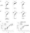
Figure 1. Developmental changes in GABA-induced currents in cerebellar granule cell GABAA receptors
A and B, membrane currents evoked by rapidly applied 0.5-100 μm GABA at 9 DIV (A) and 13 DIV (B). Current calibration of 100 pA corresponds to A and 200 pA to B. GABA was applied for the duration indicated by the continuous line. Records are from 2 neurones at pH 7.4 and recorded under whole-cell voltage clamp at -50 mV. C, left, normalised GABA-activated current (ΔIN)-concentration curves constructed at 6 DIV (○), 8 DIV (□), 9 DIV (▵), 10 DIV (⋄), 11 DIV (□), 12 DIV (•), 13 DIV (▪), 14 DIV (▴), 15 DIV (♦) and 17 DIV (⋆). Right, the same developmental data represented as two separate GABA concentration curves at 6-11 DIV (•) and 12-17 DIV (•). All data were fitted to the Hill equation (n = 30) and EC50 values and Hill coefficients determined (Table 1).
Figure 2
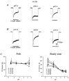
Figure 2. Proton sensitivity of cerebellar granule cell GABAA receptors at early developmental times
A and B, membrane currents activated by 10 μm GABA at 6 DIV in granule cells at -50 mV holding potential. Cells were exposed to either pH 5.4 (A) or pH 9.4 (B) Krebs solution followed by a recovery (5 min) at pH 7.4. C, pH titration for the peak (left) and steady-state (right) GABA-activated inward currents at 3 DIV (•), 6 DIV (□), 8 DIV (▵) and 10 DIV (⋄). Data are normalised to the response obtained at pH 7.4 at each developmental time. All points are means ±s.e.m. (n = 12). The data were fitted with the pH model (see Methods) and pKa values determined (Table 1).
Figure 3
Figure 3. Modulation of GABA-activated currents in cultured cerebellar granule cell neurones by H+ at late developmental times
A and B, membrane currents were activated by 2.5 μm GABA and recorded at -50 mV holding potential from granule neurones at 14 DIV. Cells were exposed to GABA at pH 5.4 (A) and pH 9.4 (B) followed by a recovery at pH 7.4. C, pH titration for the peak (left) and steady-state (right) GABA-activated currents at 12 DIV (•), 14 DIV (□), 16 DIV (▵), 17 DIV (⋄) and 20 DIV (⋆) (n = 15). The data for the steady-state GABA-induced responses were fitted with the pH model and pKa values determined (Table 1).
Figure 4
Figure 4. Voltage dependence of GABA-activated current modulation by H+
A and B, current-voltage relationships for the peak (left) and steady-state (right) GABA-activated responses at 5 DIV (A; 10 μm GABA) and 15 DIV (B; 2.5 μm GABA) at pH 7.4 (□) and pH 5.4 (•). The curves for peak and steady-state GABA-activated responses were generated using either 2nd or 3rd order polynomials. The GABA reversal potentials did not deviate significantly from 0 mV.
Figure 5
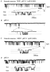
Figure 5. H+ modulation of single-channel GABA-activated currents from cerebellar granule neurones at differing developmental stages
GABA (1 μm)-activated single-channel currents recorded from 2 representative neurones at 7 DIV (A and B) and 14 DIV (C and D) at -70 mV holding potential. For the patch from a 7 DIV neurone, at pH 7.4, openings are composed of single events and bursts. A selected time period (continuous bar) has been expanded to show the different conductance levels indicated by dotted lines. In pH 5.4 Krebs solution (B), the frequency of channel opening was reduced. For the patch from a 14 DIV neurone, exposure to pH 5.4 (D) has little effect on channel frequency or conductance. These records have been filtered at 1.5 kHz for display.
Figure 6
Figure 6. Effect of development and H+ concentration on GABA single-channel conductance
Analysis of the 3 GABA single-channel conductance states, 22-26 pS (high), 16-17 pS (medium) and 12-14 pS (low), at the stages of development 4-16 DIV, following exposure to external pH 5.4, 6.4, 7.4, 8.4 and 9.4. The percentage contribution of each conductance state is indicated in the inset. The data were collated from 98 outside-out patches of cerebellar granule neurones.
Figure 7
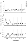
Figure 7. Effect of H+ on the mean open and shut times and Po during development
Mean open (A) and shut (B) times in addition to open probability (Po; C) were collated from single GABA channels activated by 1 μm GABA obtained from cerebellar neurones at 4-16 DIV at pH values 5.4, 6.4, 7.4, 8.4 and 9.4. Whilst mean open times are largely unaffected by pH, mean shut times, particularly at pH 5.4 and 6.4 are increased, an effect dependent upon the stage of neuronal development. The relationship of Po demonstrates a reduction at pH 5.4 and 6.4 at early development times. Data obtained from 95 outside-out patches held at -70 mV.
Figure 8
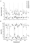
Figure 8. Effect of H+ concentration on single-channel open times during GABAA receptor development in cerebellar neurones
Single-channel open-time constants (τO1 and τO2; A) and the corresponding relative areas (AO1 and AO2; B) were measured at pH values 5.4, 6.4, 7.4, 8.4 and 9.4 at 4-16 DIV (n = 95). The time constants and relative areas were unaffected by varying extracellular H+ concentration with no obvious correlation over the developmental time period.
Figure 9
Figure 9. Effect of H+ concentration on all shut times for GABA-activated channels at different developmental stages
The 3 shut time constants (short, τC1; intermediate, τC2; and long, τC3) are plotted against the developmental stage of the cerebellar neurone and correlated with the pH of the Krebs solution. The short and intermediate shut time constants are largely unaffected by development or external pH; however, the long shut time constants are significantly greater at the lower pH values of 5.4 and 6.4 (n = 95). Patch holding potential, -70 mV.
Figure 10
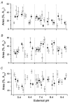
Figure 10. Relative contributions of the 3 shut time constants at various stages of development and at different H+ concentrations
The relative areas of the shut time constants corresponding to AC1C1), AC2C2) and AC3C3) were ascertained from individual shut time frequency histograms constructed at 4-16 DIV at pH 5.4, 6.4, 7.4, 8.4 and 9.4 (n = 95). Note the increased contribution from long shut times at low pH and early development times in C.

Similar articles

Cited by

References

    1. Angelotti TP, Macdonald RL. Assembly of GABAA receptor subunits: α1β1 and α1β1γ2S subunits produce simple unique ion channels with dissimilar single-channel properties. Journal of Neuroscience. 1993;13:1429–1440. - PMC - PubMed
    1. Angelotti TP, Tan F, Chahine KG, Macdonald RL. Molecular and electrophysiological characterization of an allelic variant of the rat α6 GABAA receptor subunit. Molecular Brain Research. 1992;16:173–178. - PubMed
    1. Beattie CE, Siegel RE. Developmental cues modulate GABAA receptor subunit mRNA expression in cultured cerebellar granule neurons. Journal of Neuroscience. 1993;13:1784–1792. - PMC - PubMed
    1. Bonnert TP, McKernan RM, Farrar S, Le Bourdelles B, Heavens RP, Smith DW, Hewson L, Rigby MR, Sirinathsinghji DJ, Brown N, Wafford KA, Whiting PJ. T, a novel γ-aminobutyric acid type A receptor subunit. Proceedings of the National Academy of Sciences of the USA. 1999;96:9891–9896. - PMC - PubMed
    1. Bovolin P, Santi MR, Puia G, Costa E, Grayson D. Expression pattern of γ-aminobutyric acid type A receptor subunit mRNAs in primary cultures of granule neurons and astrocytes from neonatal rat cerebella. Proceedings of the National Academy of Sciences of the USA. 1992;89:9344–9348. - PMC - PubMed

Publication types

LinkOut - more resources