. 2001 Jan 15;530(Pt 2):263-72.
doi: 10.1111/j.1469-7793.2001.0263l.x.

Cross-bridge interaction kinetics in rat myocardium are accelerated by strong binding of myosin to the thin filament

Affiliations

Cross-bridge interaction kinetics in rat myocardium are accelerated by strong binding of myosin to the thin filament

D P Fitzsimons et al. J Physiol. .

Abstract

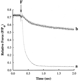
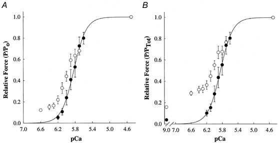
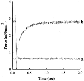
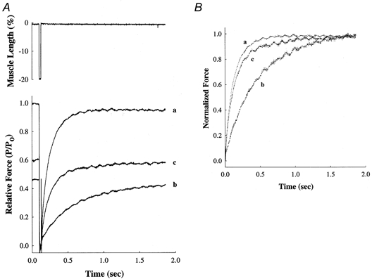
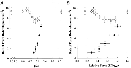
To determine the ability of strong-binding myosin cross-bridges to activate the myocardial thin filament, we examined the Ca2+ dependence of force and cross-bridge interaction kinetics at 15 degrees C in the absence and presence of a strong-binding, non-force-generating derivative of myosin subfragment-1 (NEM-S1) in chemically skinned myocardium from adult rats. Relative to control conditions, application of 6 microM NEM-S1 significantly increased Ca2+-independent tension, measured at pCa 9.0, from 0.8 +/- 0.3 to 3.7 +/- 0.8 mN mm-2. Furthermore, NEM-S1 potentiated submaximal Ca2+-activated forces and thereby increased the Ca2+ sensitivity of force, i.e. the [Ca2+] required for half-maximal activation (pCa50) increased from pCa 5.85 +/- 0.05 to 5.95 +/- 0.04 (change in pCa50 (dpCa50) = 0.11 +/- 0.02). The augmentation of submaximal force by NEM-S1 was accompanied by a marked reduction in the steepness of the force-pCa relationship for forces less than 0.50 Po (maximum Ca2+-activated force), i.e. the Hill coefficient (n2) decreased from 4.72 +/- 0.38 to 1.54 +/- 0.07. In the absence of NEM-S1, the rate of force redevelopment (ktr) was found to increase from 1.11 +/- 0.21 s-1 at submaximal [Ca2+] (pCa 6.0) to 9.28 +/- 0.41 s-1 during maximal Ca2+ activation (pCa 4.5). Addition of NEM-S1 reduced the Ca2+ dependence of ktr by eliciting maximal values at low levels of Ca2+, i.e. ktr was 9.38 +/- 0.30 s-1 at pCa 6.6 compared to 9.23 +/- 0.27 s-1 at pCa 4. At intermediate levels of Ca2+, ktr was less than maximal but was still greater than values obtained at the same pCa in the absence of NEM-S1. NEM-S1 dramatically reduced both the extent and rate of relaxation from steady-state submaximal force following flash photolysis of the caged Ca2+ chelator diazo-2. These data demonstrate that strongly bound myosin cross-bridges increase the level of thin filament activation in myocardium, which is manifested by an increase in the rate of cross-bridge attachment, potentiation of force at low levels of free Ca2+, and slowed rates of relaxation.


Figures

Figure 6
Figure 6. NEM-S1 markedly slows the rate of force relaxation
Myocardial preparations were bathed for 15 min in a pCa 9.0 solution in the absence or presence of 6 μm NEM-S1 and subsequently incubated for 2 min in loading solution containing (mm): Bes, 100; potassium propionate, 40.9; creatine phosphate, 15; dithiothreitol, 5; MgATP, 4; free Mg2+, 1; CaCl2, 0.12; and diazo-2, 0.25; pH 7.0 at 15°C. Force relaxation following flash photolysis of diazo-2 was recorded either in the absence (trace a) or in the presence of 6 μm NEM-S1 (trace b), where F refers to the time point at which the flash lamp was triggered. Each of the myocardial preparations generated ≈0.70 Po in the loading solution prior to photolysis of diazo-2.
Figure 1
Figure 1. Photomicrographs of rat skinned myocardium
Representative skinned myocardial preparation (dimensions: 430 μm w 174 μm) while relaxed (A, pCa 9.0) and during maximal activation (B, pCa 4.5). Sarcomere length in pCa 9.0 and 4.5 was 2.35 and 2.27 μm, respectively. Scale bar represents 50 μm.
Figure 3
Figure 3. Effects of 6 μm NEM-S1 on force-pCa relationships in skinned myocardium
Force-pCa relationships were determined from skinned ventricular myocardium (n = 7) in the absence (0) and presence (1) of NEM-S1. Smooth lines were generated by fitting the mean data with the equation: Prel=[Ca2+]n/(kn+[Ca2+]n), where Prel is force as a fraction of maximal Ca2+-activated force (Po) or maximal total force (PTot), n is the Hill coefficient, and k is the [Ca2+] required for half-maximal activation (i.e. pCa50). Data points are means and the error bars are s.e.m. A, Ca2+-activated force-pCa relationship in the absence (pCa50= 5.85 ± 0.05, n2= 4.72 ± 0.38) and presence of NEM-S1 (pCa50= 5.95 ± 0.04, n2= 1.54 ± 0.07). B, total force-pCa relationship in the absence (pCa50= 5.86 ± 0.05, n2= 4.50 ± 0.69) and presence of NEM-S1 (pCa50= 6.09 ± 0.05, n2= 0.93 ± 0.22). Total force is the sum of Ca2+-independent and Ca2+-activated forces at each pCa.
Figure 2
Figure 2. NEM-S1 increases Ca2+-independent force
A mechanical release-restretch manoeuvre was used to measure the rate of force redevelopment in a solution of pCa 9.0 in the absence and presence of 6 μm NEM-S1. This skinned myocardial preparation generated Ca2+-independent force of 0.05 Po in the absence of NEM-S1 in pCa 9.0 (trace a), whereas Ca2+-independent force increased to 0.22 Po following NEM-S1 treatment (trace b).
Figure 4
Figure 4. NEM-S1 potentiates submaximal force and the rate of force redevelopment
A, force records showing redevelopment of submaximal force developed by a skinned ventricular preparation at pCa 5.8 both in the absence (trace b) and in the presence of 6 μm NEM-S1 (trace c). Force in each case is scaled to the maximum force (Po) generated by the same preparation when exposed to a solution of pCa 4.5 (trace a). The upper panel shows the mechanical release-restretch manoeuvre used to measure ktr. B, to illustrate differences in the rate of force redevelopment, the force records in A are re-plotted with the peak force at each pCa scaled to 1.0.
Figure 5
Figure 5. NEM-S1 nearly eliminates the activation dependence of the rate of force redevelopment
Force redevelopment following rapid release and restretch was measured in rat skinned myocardium (n = 7) in the absence (0) and presence (1) of NEM-S1. Data points are means and the error bars are the s.e.m. A, the Ca2+ dependence of the rate constant of force redevelopment, ktr. B, the rate constant of force redevelopment (ktr) as a function of force (as a fraction of total force) measured at each pCa. Total force is the sum of Ca2+-independent and Ca2+-activated forces at each pCa.

References

    1. Adams SR, Kao JPY, Tsien RY. Biologically useful chelators that take up Ca2+ upon illumination. Journal of the American Chemical Society. 1989;111:7957–7968.
    1. Baker AJ, Figueredo VM, Keung EC, Camacho SA. Ca2+ regulates the kinetics of tension development in intact cardiac muscle. American Journal of Physiology. 1998;275:H744–750. - PubMed
    1. Brenner B. Effects of Ca2+ on cross-bridge turnover kinetics in skinned single rabbit psoas fibers: implications for regulation of muscle contraction. Proceedings of the National Academy of Sciences of the USA. 1988;85:3265–3269. - PMC - PubMed
    1. Brenner B, Eisenberg E. Rate of force generation in muscle: correlation with actomyosin ATPase activity in solution. Proceedings of the National Academy of Sciences of the USA. 1986;83:3542–3546. - PMC - PubMed
    1. Campbell K. Rate constant of muscle force redevelopment reflects cooperative activation as well as cross-bridge kinetics. Biophysical Journal. 1997;72:254–262. - PMC - PubMed

Publication types

LinkOut - more resources