

Comment
doi: 10.1073/pnas.98.2.398.
Can mosaic tumor vessels facilitate molecular diagnosis of cancer?
Affiliations
- PMID: 11209044
- PMCID: PMC33361
- DOI: 10.1073/pnas.98.2.398
Item in Clipboard
Comment
Can mosaic tumor vessels facilitate molecular diagnosis of cancer?
Proc Natl Acad Sci U S A.
.
No abstract available
Figures
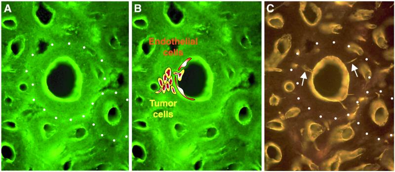
Cross-sections of breast cancer (MCa-IV) in mice showing the
microcylinders of tumor cells that surround each vessel. Large and
small thin-walled microvessels in breast tumor labeled by vascular
perfusion of green (FITC) fluorescent lectin staining
(A), or by CD31 immunoreactivity viewed by Cy3
fluorescence (gold) (C). The perivascular cuff of tumor
tissue, outlined by white dots in A and
C, is 100 microns thick, which is within the range of
the oxygen diffusion limit. In B, two endothelial cells
(red cytoplasm with white nuclei) have been drawn facing the lumen to
approximate scale. Yellow tumor cells with brown-red nuclei occupy the
perivascular cuff of tumor tissue. One tumor cell is intravasating into
the lumen and is exposed to the blood between the two endothelial
cells. This tumor cell represents the approximately one million tumor
cells per gram of tumor that may shed into the circulation each day.
The CD31 immunoreactivity, like the lectin in A and
B, defines the luminal surface of the vessels, but,
unlike the lectin, it also labels tiny sprouts (white arrows), which
have no apparent lumen because they have CD31 immunoreactivity, but no
lectin staining. These sprouts of about 1 μm diameter radiate from
the vessel lining into the 100 μm thick perivascular cuff of tumor
tissue. The sprouts result from endothelial cells that are migrating
(extravasating) from the wall of the microvessel. Vessels were
preserved in the open state by vascular perfusion of fixative (courtesy
of Donald M. McDonald, University of California, San Francisco) (2,
23). (Drawings in B by J.F. and Kristin Gullage.)
[Reproduced with permission from ref. (Copyright 2000, B. C.
Decker).]
Comment on
-
Mosaic blood vessels in tumors: frequency of cancer cells in contact with flowing blood.Proc Natl Acad Sci U S A. 2000 Dec 19;97(26):14608-13. doi: 10.1073/pnas.97.26.14608. Proc Natl Acad Sci U S A. 2000. PMID: 11121063 Free PMC article.
References
-
- Folkman J. N Engl J Med. 1971;285:1182–1186. - PubMed
-
- Folkman J. In: Cancer Medicine. 5th Ed. Holland J F, Frei E III, Bast R C Jr, Kufe D W, Pollock R E, Weichselbaum R R, editors. Ontario, Canada: B. C. Decker; 2000. pp. 132–152.
-
- Carmeleit P, Jain R K. Nature (London) 2000;407:249–257. - PubMed
-
- Lyden D, Young A Z, Zagzag D, Yan W, Gerald W, O'Reilly R, Bader B L, Hynes R O, Zhuang Y, Manova K, et al. Nature (London) 1999;401:670–677. - PubMed
Publication types
MeSH terms
Substances
LinkOut - more resources
Full Text Sources
