Comparative Study
. 2001 Jan 16;98(2):641-5.
doi: 10.1073/pnas.98.2.641.

Point mutations in the murine fumarylacetoacetate hydrolase gene: Animal models for the human genetic disorder hereditary tyrosinemia type 1

Affiliations
Comparative Study

Point mutations in the murine fumarylacetoacetate hydrolase gene: Animal models for the human genetic disorder hereditary tyrosinemia type 1

J L Aponte et al. Proc Natl Acad Sci U S A. .

Abstract

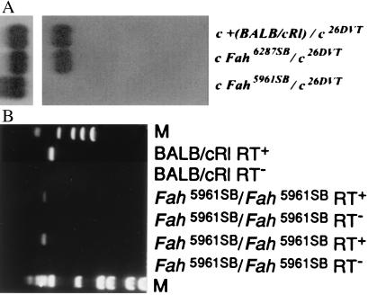
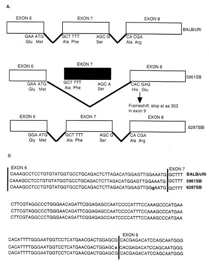
Hereditary tyrosinemia type 1 (HT1) is a severe autosomal recessive metabolic disease associated with point mutations in the human fumarylacetoacetate hydrolase (FAH) gene that disrupt tyrosine catabolism. An acute form of HT1 results in death during the first months of life because of hepatic failure, whereas a chronic form leads to gradual development of liver disease often accompanied by renal dysfunction, childhood rickets, neurological crisis, and hepatocellular carcinoma. Mice homozygous for certain chromosome 7 deletions of the albino Tyr; c locus that also include Fah die perinatally as a result of liver dysfunction and exhibit a complex syndrome characterized by structural abnormalities and alterations in gene expression in the liver and kidney. Here we report that two independent, postnatally lethal mutations induced by N-ethyl-N-nitrosourea and mapped near Tyr are alleles of Fah. The Fah(6287SB) allele is a missense mutation in exon 6, and Fah(5961SB) is a splice mutation causing loss of exon 7, a subsequent frameshift in the resulting mRNA, and a severe reduction of Fah mRNA levels. Increased levels of the diagnostic metabolite succinylacetone in the urine of the Fah(6287SB) and Fah(5961SB) mutants indicate that these mutations cause a decrease in Fah enzymatic activity. Thus, the neonatal phenotype present in both mutants is due to a deficiency in Fah caused by a point mutation, and we propose Fah(5961SB) and Fah(6287SB) as mouse models for acute and chronic forms of human HT1, respectively.


Figures

Figure 1
Figure 1
Deletion mapping of two ENU-induced postnatally lethal mutations to the Fah region of mouse chromosome 7. Horizontal lines beneath the chromosome (heaviest line) represent a subset of Tyr-locus deletions used for the mapping of the ENU-induced mutations 5961SB and 6287SB. Genetic markers are shown above the chromosome in italics. Detailed definitions and descriptions of DNA markers and loci have been published (28). The proximal breakpoints of the Del(c)14CoS and Del(c)11DSD deletions flank the Fah region. The proximal breakpoint of the Del(c)10R75 M deletion maps <1 kb 5′ to the start of Fah transcription and is believed to remove sequences essential for expression of Fah (23).
Figure 2
Figure 2
Northern blot analysis of the Fah6287SB and Fah 5961 alleles and RT-PCR analysis of the Fah 5961 allele. (A) Expression of Fah mRNA (1.4-kb transcript) in 3 μg of poly(A)+ fetal liver RNA from control [BALB/cRl/Del(c)26DVT] mice (lane 1), Fah6287SB/Del(c)26DVT (lane 2), and Fah5961SB/Del(c)26DVT (lane 3) mutants is shown. The genotype of the animals is given above; the probe for Upper is the full-length Fah cDNA and for Lower is β-actin (hybridized sequentially). (B) PCR-amplification of BALB/cRl and homozygous 5961SB mutant Fah cDNA with primers that amplify exon 7. Genotypes are given above. Fragments in lanes 2, 4, and 6 were amplified in the presence of reverse transcriptase, whereas reactions in lanes 3, 5, and 7 contained no reverse transcriptase. Lanes 1 (øX174/HaeIII) and 8 (1-kb DNA ladder) contain molecular-mass standards (M).
Figure 3
Figure 3
Schematic representation of the murine Fah6287SB and Fah5961SB point mutations and their effect on the processing of the Fah mRNA. (A) Top illustrates the normal splicing pattern for Fah followed by the altered pattern in Fah5961SB and a similar schematic for Fah6287SB. (Middle) As indicated, the Fah5961SB mutation results in an altered pattern of mRNA splicing, causing the loss of exon 7 from the Fah mRNA and a subsequent shift in the reading frame to introduce a premature stop codon in exon 9. Bottom shows the relative position of the missense mutation in Fah6287SB. (B) Nucleotide sequences of Fah exons 6, 7, and 8 of control BALB/cRl (line 1), mutant 6287SB (line 2), and mutant 5961SB (line 3). Mutant Fah6287SB has a point mutation at nucleotide 602, as indicated by the “g” in exon 6 in line 2. Mutant Fah5961SB has a point mutation in the last position of exon 7, as indicated by the “a” in line 3, that destroys the consensus splice donor sequence.

Similar articles

Cited by

References

    1. Goldsmith L A, Laberge C. The Metabolic Basis of Inherited Disease. 6th Ed. Vol. 1. New York: McGraw–Hill; 1989. pp. 547–562.
    1. Berger R, van Faassen H, Smith G P A. Clin Chim Acta. 1983;134:129–141. - PubMed
    1. Overturf K, Al-Dhalimy M, Tanguay R, Brantly M, Ou C, Finegold M, Grompe M. Nat Genet. 1996;12:266–273. - PubMed
    1. Tanguay R M, Valet J P, Lescault A, Duband J L, Laberge C, Lettre F, Plante M. Am J Hum Genet. 1990;47:308–316. - PMC - PubMed
    1. Labelle Y, Phaneuf D, Leclerc B, Tanguay R M. Hum Mol Genet. 1993;2:941–946. - PubMed

Publication types

MeSH terms