Conditional immortalization of freshly isolated human mammary fibroblasts and endothelial cells
- PMID: 11209060
- PMCID: PMC14642
- DOI: 10.1073/pnas.98.2.646
Conditional immortalization of freshly isolated human mammary fibroblasts and endothelial cells
Abstract
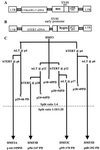
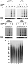
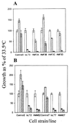
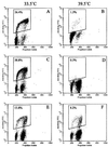
Reports differ as to whether reconstitution of telomerase activity alone is sufficient for immortalization of different types of human somatic cells or whether additional activities encoded by other "immortalizing" genes are also required. Here we show that ectopic expression of either the catalytic subunit of human telomerase (hTERT) or a temperature-sensitive mutant (U19tsA58) of simian virus 40 large-tumor antigen alone was not sufficient for immortalization of freshly isolated normal adult human mammary fibroblasts and endothelial cells. However, a combination of both genes resulted in the efficient generation of immortal cell lines irrespective of the order in which they were introduced or whether they were introduced early or late in the normal proliferative lifespan of the cultures. The order and timing of transduction, however, did influence genomic stability. Karyotype analysis indicated that introduction of both transgenes at early passage, with hTERT first, yielded diploid cell lines. Temperature-shift experiments revealed that maintenance of the immortalized state depended on continued expression of functional U19tsA58 large-tumor antigen, with hTERT alone unable to maintain growth at nonpermissive temperatures for U19tsA58 large-tumor antigen. Such conditional diploid lines may provide a useful resource for both cell engineering and for studies on immortalization and in vitro transformation.
Figures
Similar articles
-
Comparison of human mammary epithelial cells immortalized by simian virus 40 T-Antigen or by the telomerase catalytic subunit.Oncogene. 2002 Jan 3;21(1):128-39. doi: 10.1038/sj.onc.1205014. Oncogene. 2002. PMID: 11791183
-
Mass cultured human fibroblasts overexpressing hTERT encounter a growth crisis following an extended period of proliferation.Exp Cell Res. 2000 Sep 15;259(2):336-50. doi: 10.1006/excr.2000.4982. Exp Cell Res. 2000. PMID: 10964501
-
Viral oncogenes accelerate conversion to immortality of cultured conditionally immortal human mammary epithelial cells.Oncogene. 1999 Apr 1;18(13):2169-80. doi: 10.1038/sj.onc.1202523. Oncogene. 1999. PMID: 10327063
-
Senescence as a mode of tumor suppression.Environ Health Perspect. 1991 Jun;93:59-62. doi: 10.1289/ehp.919359. Environ Health Perspect. 1991. PMID: 1663451 Free PMC article. Review.
-
Immortalization of human preadipocytes.Biochimie. 2003 Dec;85(12):1231-3. doi: 10.1016/j.biochi.2003.10.015. Biochimie. 2003. PMID: 14739075 Review.
Cited by
-
Functional haplotypes of the hTERT gene, leukocyte telomere length shortening, and the risk of peripheral arterial disease.PLoS One. 2012;7(10):e47029. doi: 10.1371/journal.pone.0047029. Epub 2012 Oct 17. PLoS One. 2012. PMID: 23082138 Free PMC article.
-
Passage-dependent changes in baboon endothelial cells--relevance to in vitro aging.DNA Cell Biol. 2004 Aug;23(8):502-9. doi: 10.1089/1044549041562294. DNA Cell Biol. 2004. PMID: 15307953 Free PMC article.
-
Modeling of replicative senescence in hematopoietic development.Aging (Albany NY). 2009 Jul 23;1(8):723-32. doi: 10.18632/aging.100072. Aging (Albany NY). 2009. PMID: 20195386 Free PMC article.
-
HNRNPL Restrains miR-155 Targeting of BUB1 to Stabilize Aberrant Karyotypes of Transformed Cells in Chronic Lymphocytic Leukemia.Cancers (Basel). 2019 Apr 23;11(4):575. doi: 10.3390/cancers11040575. Cancers (Basel). 2019. PMID: 31018621 Free PMC article.
-
Mechanically tuned 3 dimensional hydrogels support human mammary fibroblast growth and viability.BMC Cell Biol. 2017 Dec 16;18(1):35. doi: 10.1186/s12860-017-0151-y. BMC Cell Biol. 2017. PMID: 29246104 Free PMC article.
References
Publication types
MeSH terms
Substances
LinkOut - more resources
Full Text Sources
Other Literature Sources
Research Materials

