. 2001 Jan 16;98(2):664-9.
doi: 10.1073/pnas.98.2.664.

Bunyamwera bunyavirus nonstructural protein NSs is a nonessential gene product that contributes to viral pathogenesis

Affiliations

Bunyamwera bunyavirus nonstructural protein NSs is a nonessential gene product that contributes to viral pathogenesis

A Bridgen et al. Proc Natl Acad Sci U S A. .

Abstract

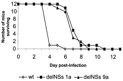
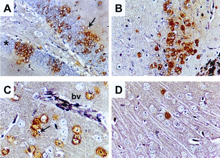
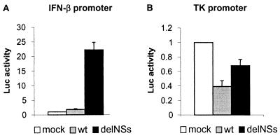
Bunyamwera virus (family Bunyaviridae, genus Bunyavirus) contains a tripartite negative-sense RNA genome. The smallest RNA segment, S, encodes the nucleocapsid protein N and a nonstructural protein, NSs, in overlapping reading frames. We have generated a mutant virus lacking NSs, called BUNdelNSs, by reverse genetics. Compared with the wild-type (wt) virus, BUNdelNSs exhibited a smaller plaque size and generated titers of virus approximately 1 log lower. In mammalian cells, the mutant expressed greatly increased levels of N protein; significantly, the marked inhibition of host cell protein synthesis shown by wt virus was considerably impaired by BUNdelNSs. When inoculated by the intracerebral route BUNdelNSs killed BALB/c mice with a slower time course than wt and exhibited a reduced cell-to-cell spread, and titers of virus in the brain were lower. In addition, the abrogation of NSs expression changed Bunyamwera virus from a noninducer to an inducer of an interferon-beta promoter. These results suggest that, although not essential for growth in tissue culture or in mice, the bunyavirus NSs protein has several functions in the virus life cycle and contributes to viral pathogenesis.


Figures

Figure 1
Figure 1
Nucleotide sequences from bases 86 to 122 of BUN S segment in pT7riboBUNS, pT7riboBUNN, and pT7riboBUNNSs showing the N and NSs start codons and mutations made to ablate either the N or NSs reading frames. Amino acid translations are given below the nucleotide sequences. Initiating methionine residues are indicated in bold lettering and stop codons by an asterisk.
Figure 2
Figure 2
Comparison of plaques produced by wt BUN (Left) and the BUNdelNSs mutant (Right) in BHK cells. Cell monolayers were fixed with 4% formaldehyde and stained with Giemsa's solution.
Figure 3
Figure 3
Characterization of the transfectant virus genome. (A) Northern blot analysis. BHK cells were infected with wt BUN (lane 1) or with transfectant viruses 1a (lane 2) or 9a (lane 3). Total cell RNA was extracted at 31 h after infection and separated on a 1.2% agarose gel containing 3.7% formaldehyde. Blotted RNA was hybridized with a mixture of 32P-labeled riboprobes that detected the L, M, and S RNA segments, as indicated. (B) RT-PCR analysis. Virion RNA was reverse transcribed by using primer NS2, and cDNA products were amplified by PCR by using primers S4 and NS2. PCR products from plasmid constructs pT7riboBUNN and pT7riboBUNNSs are included as markers for the expected product sizes. Lane 1, 1-kbp ladder; lane 2, PCR negative control; lane 3, PCR product from pT7riboBUNNSs; lane 4, PCR product from pT7riboBUNN; lane 5, RT-PCR product from transfectant virus 1a RNA; lane 6, RT-PCR product from transfectant virus 9a RNA. (C) Nucleotide sequence comparison. The nucleotide sequences of cloned RT-PCR products from the S segment RNA of wt BUN (Left) or transfectant virus 9a (Right) were determined in the region of the NSs start codon. The nucleotide changes from the wt to the mutant sequence are indicated (Right).
Figure 4
Figure 4
Protein profiles of wt and transfectant bunyaviruses. (A) Absence of NSs protein and overexpression of N protein in cells infected with the transfectant viruses. BHK cells were infected with wt BUN (lane 1), Maguari virus (lane 2), or with transfectant viruses BUNdelNSs 1a (lane 3) or BUNdelNSs 9a (lane 4) and labeled with [35S]methionine for 2 h at 24 h after infection. Cell lysates were separated by SDS/PAGE, and labeled proteins were visualized by autoradiography. Positions of bunyavirus proteins are indicated. (B) Shutoff of host cell protein synthesis. BHK cells were infected with 5 pfu per cell of wt BUN (lane 1), transfectant virus BUNdelNSs 1a (lane 2), and transfectant virus BUNdelNSs 9a (lane 3) or were mock infected (lane 4). Cells were labeled with [35S]methionine for 2 h at 48 h after infection, and then equal amounts of cell lysate were analyzed by SDS/PAGE.
Figure 5
Figure 5
Pathogenesis of bunyaviruses in mice. Three groups of 12 5-week-old female BALB/c mice were inoculated i.c. with 2,000 pfu of wt BUN or BUNdelNSs viruses 1a or 9a. The animals were monitored for 14 days. Any mice that were moribund or severely paralyzed were killed and scored as dead that day.
Figure 6
Figure 6
Histology and immunostaining of brain sections from infected BALB/c mice. Animals were infected with 2,000 pfu of wt BUN (A–C) or BUNdelNSs 1a (D) by the intracerebral route. Mice were killed 4 days after infection, and brain sections were prepared. Micrographs show different areas of the brain immunostained for viral proteins (brown). (A) Foci of positive neurones in the hippocampal dentate gyrus. Note that staining is predominantly cytoplasmic and that several infected cells have enlarged pale staining nuclei (arrow). For comparison, unstained neurones with a normal morphology are indicated by an asterisk. (B) Positive neurones in the anterior olfactory nucleus. C and D show sections of the frontal cortex. (C) Positive neurones adjacent to a blood vessel (bv) in the cortex. Note again the enlarged pale staining nuclei (arrow) of infected cells. (D) Infection with BUNdelNSs virus results in fewer infected cells.
Figure 7
Figure 7
Induction of an IFN-β promoter by infection with BUNdelNSs virus. Monolayers of BF cells were transfected in parallel with the IFN-β promoter-containing plasmid (A) or with the herpes simplex virus-thymidine kinase promoter-containing control plasmid (B). At 6 h after transfection, the cells were mock infected or infected with wt BUN or BUNdelNSs virus 9a at 5 pfu per cell. Fifteen hours after infection, luciferase activities in cell lysates were measured. The activity measured in mock-infected cells was taken as 1, and averages (columns) and standard deviations (error bars) of the luciferase activities of three independent experiments are shown.

Similar articles

Cited by

References

    1. Elliott R M. Mol Med. 1997;3:572–577. - PMC - PubMed
    1. Bishop D H L. In: The Bunyaviridae. Elliott R M, editor. New York: Plenum; 1996. pp. 19–61.
    1. Bouloy M. Adv Virus Res. 1991;40:235–275. - PubMed
    1. Elliott R M. J Gen Virol. 1990;71:501–522. - PubMed
    1. Schmaljohn C S. In: Virology. 2nd Ed. Fields B N, Knipe D M, editors. New York: Raven; 1996. pp. 1447–1471.

Publication types

MeSH terms

LinkOut - more resources