Comparative Study
. 2001 Jan 16;98(2):670-5.
doi: 10.1073/pnas.98.2.670.

Antigenic and genetic heterogeneity of Borrelia burgdorferi populations transmitted by ticks

Affiliations
Comparative Study

Antigenic and genetic heterogeneity of Borrelia burgdorferi populations transmitted by ticks

J Ohnishi et al. Proc Natl Acad Sci U S A. .

Abstract

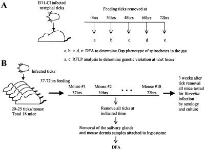
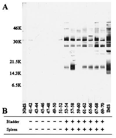
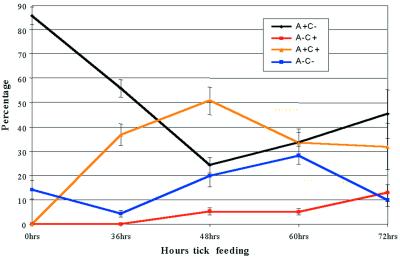
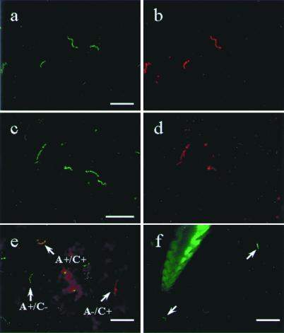
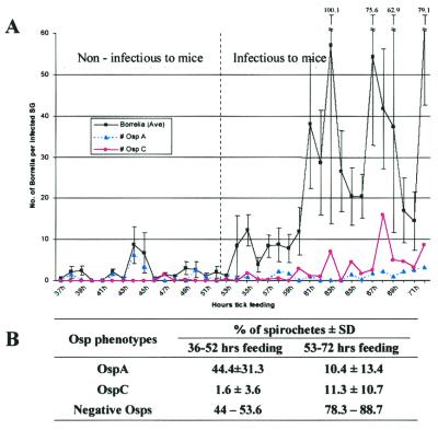
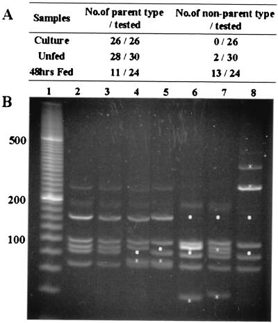
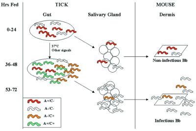
The genome of Borrelia burgdorferi encodes a large number of lipoproteins, many of which are expressed only at certain stages of the spirochete's life cycle. In the current study we describe the B. burgdorferi population structure with respect to the production of two lipoproteins [outer surface protein A (OspA) and outer surface protein C (OspC)] during transmission from the tick vector to the mammalian host. Before the blood meal, the bacteria in the tick were a homogeneous population that mainly produced OspA only. During the blood meal, the population became more heterogeneous; many bacteria produced both OspA and OspC, whereas others produced only a single Osp and a few produced neither Osp. From the heterogeneous spirochetal population in the gut, a subset depleted of OspA entered the salivary glands and stably infected the host at time points >53 hr into the blood meal. We also examined genetic heterogeneity at the B. burgdorferi vlsE locus before and during the blood meal. In unfed ticks, the vlsE locus was stable and one predominant and two minor alleles were detected. During the blood meal, multiple vlsE alleles were observed in the tick. Tick feeding may increase recombination at the vlsE locus or selectively amplify rare vlsE alleles present in unfed ticks. On the basis of our data we propose a model, which is different from the established model for B. burgdorferi transmission. Implicit in our model is the concept that tick transmission converts a homogeneous spirochete population into a heterogeneous population that is poised to infect the mammalian host.


Figures

Figure 1
Figure 1
Outline of experimental approach. (A) B. burgdorferi Osp phenotype in the tick gut. Infected ticks were placed on mice and removed at the indicated times. Twelve unfed and 93 fed nymphal ticks (12 ticks from 36 hr, 39 ticks from 48 hr, 36 ticks from 60 hr, and 6 ticks from 72 hr) were removed. The guts were dissected and examined by DFA to determine the OspA and OspC phenotype within the gut. DNA was purified from gut homogenates prepared from unfed and partially engorged (48-hr) ticks to determine the extent of variation at the B. burgdorferi vlsE locus. (B) B. burgdorferi infection of tick salivary glands and mouse dermis. A total of 18 mice were infested with 20–25 infected nymphal ticks. All of the ticks were removed from individual mice at 2-hr intervals starting at 37 hr and ending at 72 hr into the blood meal. Mouse and tick samples were analyzed for B. burgdorferi infection and Osp phenotypes.
Figure 2
Figure 2
Timing of tick transmission of B. burgdorferi to mice. Mice exposed to infected ticks for various time periods (Fig. 1B) were tested for infection by Western blotting (A) or culture (B). Numbers on the left in A are molecular masses of markers in kDa. In a few cases (hr 37–38, 39–40, 55–56) all of the ticks were not recovered from the mice. These animals were not tested for infection because we could not accurately determine the length of tick exposure. IMS, infected mouse serum; NMS, normal (uninfected) mouse serum.
Figure 3
Figure 3
Population dynamics of B. burgdorferi producing OspA and OspC in the tick gut during the blood meal. Infected ticks were allowed to feed on mice for various time periods as described for Fig. 1A. The guts were removed from ticks and the proportion of bacteria displaying each Osp phenotype was determined as described in the text.
Figure 4
Figure 4
Direct dual immunofluorescence of B. burgdorferi in the gut of partially engorged (48 hr) ticks and in the mouse dermis. (a and b) The same field double-labeled with a FITC-conjugated polyclonal B. burgdorferi antibody (a) and a TR-conjugated mAb against OspA (b). (c and d) The same field labeled with FITC-conjugated polyclonal Borrelia antibody (c) and a TR-conjugated mAb against OspC (d). (e) Merged image of spirochetes stained with Alexa 488-conjugated OspA (green) and TR-conjugated OspC (red). Three phenotypes were observed in e: arrows, Borrelia producing only OspA in green, only OspC in red, and both OspA and OspC in orange. (f) Mouse skin sample attached to the hypostome of a tick that had fed for 40 hr is labeled with the FITC-conjugated polyclonal Borrelia antibody. The hypostome itself autofluoresces, and bacteria staining with the antibody are indicated by the arrowhead. (Bars in a, c, and e represent 20 μm, and the bar in f represents 50 μm.)
Figure 5
Figure 5
B. burgdorferi infection of tick salivary glands. (A) Salivary glands were removed from ticks attached to mice for various time periods as described for Fig. 1B. At each time point the samples were analyzed to determine the mean number of total spirochetes per infected salivary gland (SG) pair (♦) and the mean number of OspA- (▴) and OspC- (●) producing spirochetes per infected pair of glands. The vertical dashed line separates ticks that had attached to the host for too short a time to infect mice (<53 hr) from those that had attached long enough to infect mice (≥53 hr) (see Fig. 2 for mouse transmission data). (B) Summary of Osp phenotypes in the salivary glands before and after mouse infection during tick feeding. The salivary gland data are based on A.
Figure 6
Figure 6
RFLP analysis of the vlsE locus of B. burgdorferi in culture, unfed, and partially engorged (48-hr) ticks. (A) Summary of the vlsE variants detected by RFLP analysis. The single vlsE allele in cultured bacteria was designated as the parental allele. In partially fed ticks, the 24 clones belonged to 13 different RFLP types, all of which were different from the parent type. (B) Lane 1, 20-bp ladder; lane 2, RFLP pattern of parental B31-C1; lanes 3 and 4, RFLP patterns of 2 of the 3 alleles detected in unfed ticks; lanes 5–9, RFLP patterns of 4 of the 13 alleles detected in fed ticks. The asterisks mark new bands in the clones that are absent from the parental strain. The dots mark bands present in parental strain that are absent from the clone tested.
Figure 7
Figure 7
Model for population dynamics of OspA and OspC during B. burgdorferi (Bb) transmission from ticks to mice. The model is based on the predominant Osp phenotypes observed in the different organs. See text for details of model. In brief, we propose that at early times of feeding (<53 hr) noninfectious bacteria that mainly produce OspA and others that produce neither Osp invade the salivary glands and host dermis in small numbers, but they fail to stably infect the mice. Stable transmission is initiated later (≥53 hr) when bacteria producing only OspC leave the gut. These infectious organisms clear OspC en route to the salivary glands and then enter the host dermis. In our model, from the heterogeneous population in the gut, only a subpopulation that is depleted of OspA is successfully transmitted to mice.

Similar articles

Cited by

References

    1. Piesman J. Exp Appl Acarol. 1989;7:71–80. - PubMed
    1. Burgdorfer W, Lane R S, Barbour A G, Gresbrink R A, Anderson J R. Am J Trop Med Hyg. 1985;34:925–930. - PubMed
    1. Benach J L, Coleman J L, Skinner R A, Bosler E M. J Infect Dis. 1987;155:1300–1306. - PubMed
    1. de Silva A M, Telford S R, 3rd, Brunet L R, Barthold S W, Fikrig E. J Exp Med. 1996;183:271–275. - PMC - PubMed
    1. Piesman J. J Infect Dis. 1993;167:1082–1085. - PubMed

Publication types

MeSH terms