

Emotion-induced changes in human medial prefrontal cortex: II. During anticipatory anxiety
- PMID: 11209066
- PMCID: PMC14649
- DOI: 10.1073/pnas.98.2.688
Emotion-induced changes in human medial prefrontal cortex: II. During anticipatory anxiety
Abstract
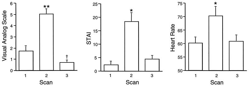
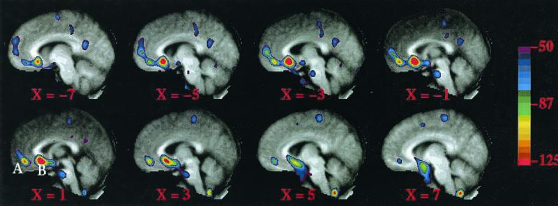
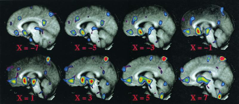
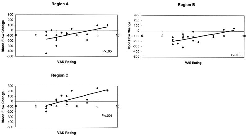
Regional cerebral blood flow (BF) was examined in the human medial prefrontal cortex (MPFC) with positron emission tomography during anticipatory anxiety. Transient anxiety was induced in normal subjects by having them anticipate a painful shock to the fingers of one hand. BF was decreased during anticipatory anxiety, relative to an eyes-closed resting condition, in two regions of the MPFC (Brodmann Areas 10/32 and 24/25). BF decreases in these areas were inversely correlated with anxiety self rating, such that the least anxious subjects exhibited the largest BF reductions, whereas the most anxious subjects showed no significant BF reduction or a slight increase. BF changes in MPFC and in the midbrain were correlated with each other and with anxiety self rating. These results are consistent with the hypothesis that BF reductions in MPFC, previously observed in cognitive tasks, reflect a dynamic balance between focused attention and subject anxiety and may occur from a functionally active baseline or default state. The characterization of such relationships within the human brain enables new insights into the integration of cognition and emotion.
Figures
References
-
- Shulman G L, Fiez J A, Corbetta M, Buckner R L, Miezin F M, Raichle M E, Petersen S E. J Cognit Neurosci. 1997;9:648–663. - PubMed
-
- Hutchinson M, Schiffer W, Joseffer S, Liu A, Schlosser R, Dikshit S, Goldberg E, Brodie J D. Magn Reson Imaging. 1999;17:1427–1436. - PubMed
-
- Eslinger P J, Damasio A R. Neurology. 1985;35:1731–1741. - PubMed
Publication types
MeSH terms
Grants and funding
LinkOut - more resources
Full Text Sources
Other Literature Sources
Medical
Miscellaneous
