. 2001 Jan 16;98(2):717-22.
doi: 10.1073/pnas.98.2.717.

Estradiol rapidly inhibits soluble guanylyl cyclase expression in rat uterus

Collaborators, Affiliations

Estradiol rapidly inhibits soluble guanylyl cyclase expression in rat uterus

J S Krumenacker et al. Proc Natl Acad Sci U S A. .

Abstract

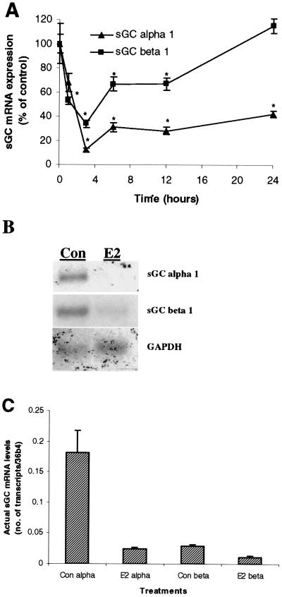
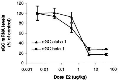
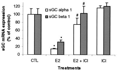
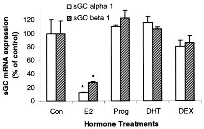
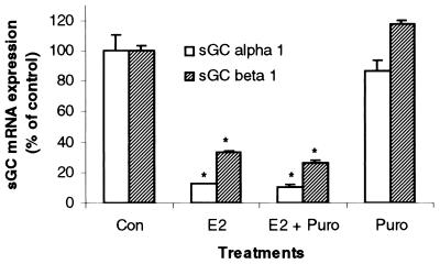
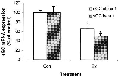
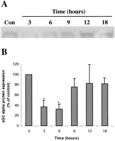
Previous reports that investigated the regulation of the NO/soluble guanylyl cyclase (sGC)/cGMP pathway by estrogenic compounds have focused primarily on the levels of NO, NO-producing enzymes, and cGMP in various tissues. In this study, we demonstrate that 17beta-estradiol (E2) regulates the alpha(1) and beta(1) subunits of the NO receptor, sGC, at the mRNA and protein levels in rat uterus. Using real-time quantitative PCR, we found that within 1 h of in vivo E2 administration to rats, sGC mRNA levels begin to diminish. After 3 h, there is a maximal diminution of sGC mRNA expression (sGC alpha(1) 10% and sGC beta(1) 33% of untreated). This effect was blocked by the estrogen receptor antagonist, ICI 182,780, indicating that estrogen receptor is required. The effect of E2 also was observed in vitro with incubations of uterine tissue, indicating that the response does not depend on the secondary release of other hormones or factors from other tissues. Puromycin did not block the effect, suggesting the effects occur because of preexisting factors in uterine tissues and do not require new protein synthesis. Using immunoblot analysis, we found that sGC protein levels also were reduced by E2 over a similar time course as the sGC mRNA. We conclude that sGC plays a vital role in the NO/sGC/cGMP regulatory pathway during conditions of elevated estrogen levels in the rat uterus as a result of the reduction of sGC expression.


Figures

Figure 1
Figure 1
E2 rapidly diminishes sGC α1 and β1 mRNA expression in the rat uterus. Ovariectomized rats were administered E2 (40 μg/kg body weight, single injection) and killed over a time course. Uterine sGC α1 and β1 mRNA expression was determined by real-time Q-PCR (0–24 h) (A) and Northern blot (at 3 h) (B). Mean values ± SEM (n = 3) in A were normalized to 36b4 and graphed as percent of control (Con, Time 0 h). (C) The actual quantitation of sGC α1 and β1 transcript levels relative to 36b4 transcript under E2-stimulated and nonstimulated conditions as determined by real-time Q-PCR.
Figure 2
Figure 2
Dose-dependent reduction of sGC α1 and β1 transcripts by E2. Increasing doses of E2 were administered to ovariectomized animals that were killed after 3 h. Uterine sGC mRNA levels were determined by real-time Q-PCR. Mean values ± SEM (Con, n = 6; all doses, n = 3) were normalized to 36b4 and graphed as percent of control.
Figure 3
Figure 3
ICI 182,780 blocks the E2-mediated diminution of uterine sGC mRNA expression. Ovariectomized rats were treated with the pure ER antagonist, ICI 182,780, 30 min before the administration of E2 (40 μg/kg body weight). Animals were killed after 3 h, and sGC α1 and β1 mRNA levels were determined by real-time Q-PCR. Mean values ± SEM (Con, n = 6; E2, n = 9; E2 + ICI, n = 6; ICI, n = 8) were normalized to 36b4 and graphed as percent of control.
Figure 4
Figure 4
Uterine sGC mRNA regulation exhibits steroid-hormone specificity. Ovariectomized animals were administered either E2 (40 μg/kg), progesterone (Prog, 40 mg/kg), 5α-dihydrotestosterone (DHT, 400 μg/kg), or dexamethasone (DEX, 600 μg/kg). Animals then were killed after 3 h and uterine sGC α1 and β1 mRNA levels were determined by real-time Q-PCR. Mean values ± SEM (n = 3) were normalized to 36b4 and graphed as percent of control.
Figure 5
Figure 5
E2 inhibition of uterine sGC mRNA levels occurs independently of new protein synthesis. Ovariectomized rats were pretreated with puromycin (30 min) and subsequently administered E2 (40 μg/kg). Animals were killed 3 h after the administration of E2, and sGC α1 and β1 mRNA levels were determined by real-time Q-PCR. Mean values ± SEM (n = 3) were normalized to 36b4 and graphed as percent of control.
Figure 6
Figure 6
E2 inhibits uterine sGC mRNA levels in vitro. Uteri were excised from ovariectomized animals and incubated in vitro for 3 h in the presence or absence of E2 (10 nM). Transcript levels for sGC α1 and β1 mRNA were determined by real-time Q-PCR. Mean values ± SEM (n = 4) were normalized to 36b4 and graphed as percent of control.
Figure 7
Figure 7
E2 leads to the reduction of sGC protein levels in rat uterus. Nonovariectomized rats were administered E2 (40 μg/kg) and killed over a time course. Uterine sGC α protein levels were determined by immunoblotting. (A) A representative blot from one experiment. (B) The average densitometric values from five independent experiments ± SEM graphed as percent of control.

References

    1. Yallampalli C, Byam-Smith M, Nelson S O, Garfield R E. Endocrinology. 1994;134:1971–1974. - PubMed
    1. Brenner R M, Slayden O D. In: The Physiology of Reproduction. Knobil E, Neill J D, editors. New York: Raven; 1994. pp. 541–569.
    1. Burger H G. Horm Res. 2000;53:25–29. - PubMed
    1. Hyder S M, Kirkland J L, Loose-Mitchell D S, Makela S, Stancel G M. In: Endocrine Disruptors. Naz R K, editor. Boca Raton, FL: CRC; 1999. pp. 166–186.
    1. Weiner C P, Lizasoain I, Baylis S A, Knowles R G, Charles I G, Moncada S. Proc Natl Acad Sci USA. 1994;91:5212–5216. - PMC - PubMed

Publication types

MeSH terms

LinkOut - more resources