. 2001 Mar;183(6):1945-53.
doi: 10.1128/JB.183.6.1945-1953.2001.

Cellulosomal scaffoldin-like proteins from Ruminococcus flavefaciens

Affiliations

Cellulosomal scaffoldin-like proteins from Ruminococcus flavefaciens

S Y Ding et al. J Bacteriol. 2001 Mar.

Abstract

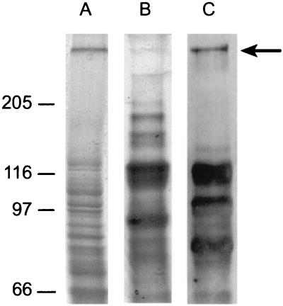
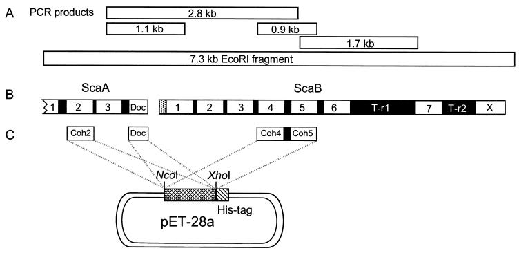
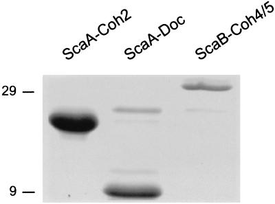
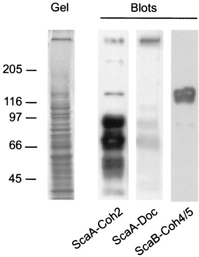
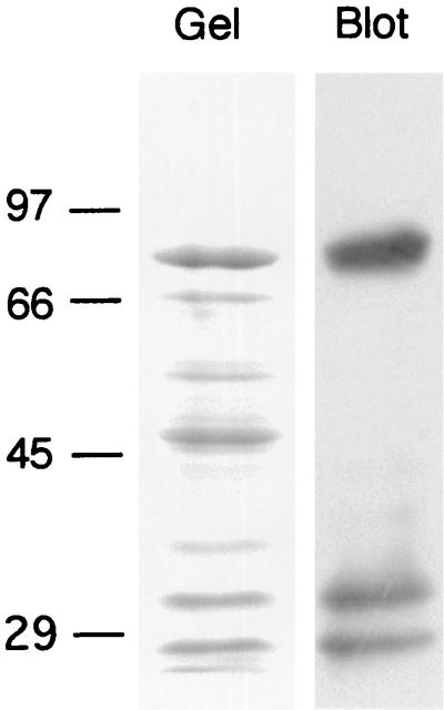
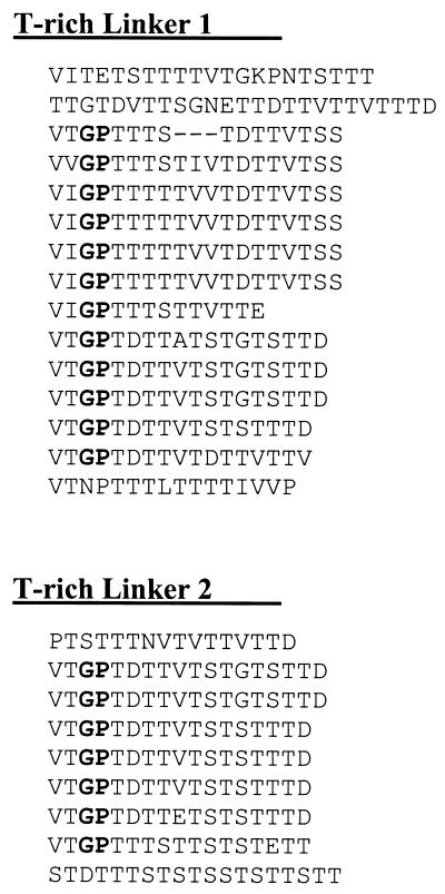
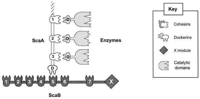
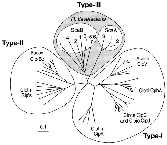
Two tandem cellulosome-associated genes were identified in the cellulolytic rumen bacterium, Ruminococcus flavefaciens. The deduced gene products represent multimodular scaffoldin-related proteins (termed ScaA and ScaB), both of which include several copies of explicit cellulosome signature sequences. The scaB gene was completely sequenced, and its upstream neighbor scaA was partially sequenced. The sequenced portion of scaA contains repeating cohesin modules and a C-terminal dockerin domain. ScaB contains seven relatively divergent cohesin modules, two extremely long T-rich linkers, and a C-terminal domain of unknown function. Collectively, the cohesins of ScaA and ScaB are phylogenetically distinct from the previously described type I and type II cohesins, and we propose that they define a new group, which we designated here type III cohesins. Selected modules from both genes were overexpressed in Escherichia coli, and the recombinant proteins were used as probes in affinity-blotting experiments. The results strongly indicate that ScaA serves as a cellulosomal scaffoldin-like protein for several R. flavefaciens enzymes. The data are supported by the direct interaction of a recombinant ScaA cohesin with an expressed dockerin-containing enzyme construct from the same bacterium. The evidence also demonstrates that the ScaA dockerin binds to a specialized cohesin(s) on ScaB, suggesting that ScaB may act as an anchoring protein, linked either directly or indirectly to the bacterial cell surface. This study is the first direct demonstration in a cellulolytic rumen bacterium of a cellulosome system, mediated by distinctive cohesin-dockerin interactions.


Figures

FIG. 1
FIG. 1
Identification of putative cellulosome-related glycoproteins from R. flavefaciens 17. A cellulose-grown culture was centrifuged, and both pellet (lane A) and supernatant fluids (lane B) were analyzed by SDS-PAGE on the same gel (7.5% polyacrylamide separating gel). A duplicate sample of the supernatant fluids was blotted onto a PVDF membrane and stained for the presence of glycosylated proteins (lane C). Molecular mass markers (in kilodaltons) are shown to the left of the gel.
FIG. 2
FIG. 2
Overview of the 7.3-kb fragment selected from an R. flavefaciens EcoRI∗ library. (A) DNA fragments crucial for solving the sequence. (B) Domain organization of the C-terminal portion of scaA and the complete scaB gene. Both genes contain multiple copies of cohesin domains (numbered), and scaA contains a C-terminal dockerin domain (Doc). All of the domains are separated by short linker sequences (black); ScaB also includes two long T-rich linking segments (T-r1 and T-r2). The sequence of scaB shows a typical signal peptide at its N terminus and a C-terminal module (X) of unknown function. (C) Overexpression of selected modules. The C-terminal cohesin 3 and dockerin domains from ScaA were expressed separately, together with a His tag, in the pET-28a vector. A double-cohesin segment (cohesins 4 and 5) from ScaB was similarly expressed.
FIG. 3
FIG. 3
SDS-PAGE of purified recombinant proteins, derived from R. flavefaciens ScaA and ScaB. Cohesin 2, the dockerin from ScaA, and the double cohesin (cohesins 4 and 5) from ScaB were subcloned and expressed as indicated in Fig. 2C. The His-tagged proteins were isolated on an Ni-nitrilotriacetic acid column; the eluted proteins were subjected to SDS-PAGE and stained with Coomassie brilliant blue. Molecular mass markers (in kilodaltons) are shown to the left of the gel.
FIG. 4
FIG. 4
Affinity blotting of cell-derived material from R. flavefaciens, using selected recombinant protein domains from ScaA and ScaB. Samples containing the pelleted, cellulose-grown culture (Fig. 1) were subjected to SDS-PAGE (Gel), and blotted onto PVDF membranes (Blots). The blots were probed with the indicated recombinant protein sample, and labeled bands (labeled at left [in kilodaltons]) were detected by chemiluminescence using peroxidase-conjugated, anti-His tag antibody.
FIG. 5
FIG. 5
Affinity blotting of a known R. flavefaciens enzyme construct with a recombinant ScaA cohesin. A GST-XynD fusion protein (molecular weight, 78,953) was subjected to SDS-PAGE (Gel), and the proteins were transferred to a PVDF membrane (Blot), and probed with ScaA-Coh2 as described in the legend to Fig. 4. Molecular mass markers (in kilodaltons) are shown to the left of the gel.
FIG. 6
FIG. 6
Alignment of internal segments within the T-rich linkers of ScaB. Note the high degree of internal repetition, the marked similarity among segments of these two linker regions, and the presence of distinctive Gly-Pro dyads (shown in boldface type). T-r1 comprises residues 939 to 1213, and T-r2 comprises residues 1356 to 1511.
FIG. 7
FIG. 7
Schematic representation of the proposed binding specificity of cellulosomal components from R. flavefaciens.
FIG. 8
FIG. 8
Phylogenetic relationship of R. flavefaciens cohesin domains. The individual ScaA and ScaB cohesins are numbered as they appear in sequence from the N terminus of the gene. The type I cohesins include those from the known scaffoldins (CipA from C. thermocellum, C. cellulolyticum CipC, C. josui CipJ, C. cellulovorans CpbA, and A. cellulolyticus CipV). Type II cohesins include those from the B. cellulosolvens CipBc scaffoldin and the surface anchoring proteins (Slp's) of C. thermocellum. The sources of the sequences used in this figure are given in reference . The scale bar indicates percentage (0.1) of amino acid substitutions.

References

    1. Aurilia V, Martin J C, McCrae S I, Scott K P, Rincon M T, Flint H J. Three multidomain esterases from the cellulolytic rumen anaerobe Ruminococcus flavefaciens 17 that carry divergent dockerin sequences. Microbiology. 2000;146:1391–1397. - PubMed
    1. Bayer E A, Chanzy H, Lamed R, Shoham Y. Cellulose, cellulases and cellulosomes. Curr Opin Struct Biol. 1998;8:548–557. - PubMed
    1. Bayer E A, Coutinho P M, Henrissat B. Cellulosome-like sequences in Archaeoglobus fulgidus: an enigmatic vestige of cohesin and dockerin domains. FEBS Lett. 1999;463:277–280. - PubMed
    1. Bayer E A, Ding S-Y, Mechaly A, Shoham Y, Lamed R. Emerging phylogenetics of cellulosome structure. In: Gilbert H J, Davies G J, Henrissat B, Svensson B, editors. Recent advances in carbohydrate bioengineering. Cambridge, United Kingdom: The Royal Society of Chemistry; 1999. pp. 189–201.
    1. Bayer E A, Morag E, Lamed R. The cellulosome—a treasure-trove for biotechnology. Trends Biotechnol. 1994;12:378–386. - PubMed

Publication types

MeSH terms

Associated data

LinkOut - more resources