. 2001 Mar;183(6):1961-73.
doi: 10.1128/JB.183.6.1961-1973.2001.

Essential thioredoxin-dependent peroxiredoxin system from Helicobacter pylori: genetic and kinetic characterization

Affiliations

Essential thioredoxin-dependent peroxiredoxin system from Helicobacter pylori: genetic and kinetic characterization

L M Baker et al. J Bacteriol. 2001 Mar.

Abstract

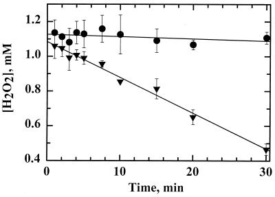
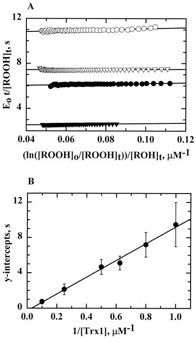
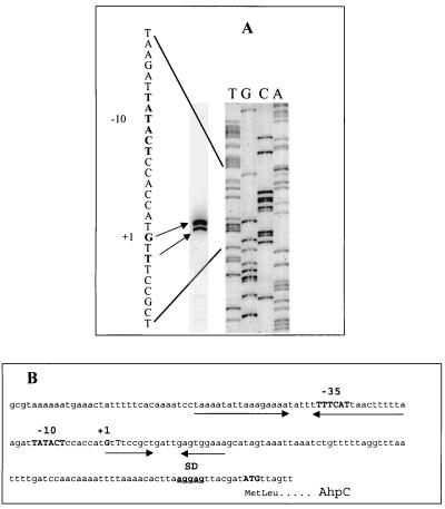
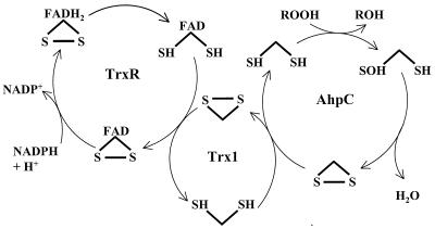
Helicobacter pylori, an oxygen-sensitive microaerophile, contains an alkyl hydroperoxide reductase homologue (AhpC, HP1563) that is more closely related to 2-Cys peroxiredoxins of higher organisms than to most other eubacterial AhpC proteins. Allelic replacement mutagenesis revealed ahpC to be essential, suggesting a critical role for AhpC in defending H. pylori against oxygen toxicity. Characterization of the ahpC promoter region divulged two putative regulatory elements and identified the transcription initiation site, which was mapped to 96 and 94 bp upstream of the initiation codon. No homologue of ahpF, which encodes the dedicated AhpC reductase in most eubacteria, was found in the H. pylori genome. Instead, homologues of Escherichia coli thioredoxin (Trx) reductase (TrxR, HP0825) and Trx (Trx1, HP0824) formed a reductase system for H. pylori AhpC. A second Trx homologue (Trx2, HP1458) was identified but was incapable of AhpC reduction, although Trx2 exhibited disulfide reductase activity with other substrates [insulin and 5,5'-dithiobis(2-nitrobenzoic acid)]. AhpC interactions with each substrate, Trx1 and hydroperoxide, were bimolecular and nonsaturable (infinite V(max) and K(m) values) but rapid enough (at 1 x 10(5) to 2 x 10(5) M(-1) s(-1)) to suggest an important role for AhpC in cellular peroxide metabolism. AhpC also exhibited a wide specificity for hydroperoxide substrates, which, taken together with the above results, suggests a minimal binding site for hydroperoxides composed of little more than the cysteinyl (Cys49) active site. H. pylori AhpC was not reduced by Salmonella typhimurium AhpF and was slightly more active with E. coli TrxR and Trx1 than was S. typhimurium AhpC, demonstrating the specialized catalytic properties of this peroxiredoxin.


Figures

FIG. 1
FIG. 1
SDS-PAGE analysis of purified AhpC, Trx1, Trx2, and TrxR from H. pylori. (A) Crude lysates and recombinant purified AhpC and TrxR were analyzed on the same 12% polyacrylamide gel in reducing sample buffer (except where noted) as follows: lane 1, molecular mass markers (Broad Range Molecular Weight Standards; Bio-Rad); lane 2, crude extracts of E. coli cells transformed with pPROK1/ahpC and induced with 0.4 mM IPTG for at least 3 h at 37°C; lane 3, pure, recombinant AhpC; lane 4, crude extracts of E. coli cells transformed with pPROK1/trxR and induced as described above; lane 5, pure, recombinant TrxR protein; lane 6, pure AhpC in nonreducing sample buffer; lane 7, pure TrxR in nonreducing sample buffer. (B) Crude lysates and recombinant Trx1 and Trx2 were analyzed on a 10% Tris-Tricine gel as follows: lane 1, molecular mass markers; lane 2, crude extracts of E. coli cells transformed with pPROK1/trx1 after induction with IPTG; lane 3, pure, recombinant Trx1; lane 4, crude extracts of E. coli cells transformed with pPROK1/trx2 after induction with IPTG; lane 5, recombinant purified Trx2 after purification. Equivalent protein masses (10 μg) were loaded in all lanes of both gels. Molecular masses (in kilodaltons) are indicated on the left.
FIG. 2
FIG. 2
Comparative kinetic parameters of Trx1 and Trx2 from H. pylori. Trx activity assays using either 200 μM DTNB (black bars) or 80 μM insulin (gray bars) as a substrate for 0 to 50 μM Trx1 or Trx2 were conducted in a standard buffer of 100 mM potassium phosphate and 2 mM EDTA, pH 7.4, with 150 μM NADPH. Assays were started with the addition of 7.0 nM TrxR and were monitored at 340 nm when insulin was used as the substrate or at 412 nm when DTNB was used as the substrate to observe the release of TNB2−. Rates were determined from the first 10% of the reaction and then fitted to a hyperbola to determine the Vmax(app) and Km(app) values for each.
FIG. 3
FIG. 3
Steady-state kinetic analysis of Trx1 and TrxR from H. pylori. Assays were conducted using a stopped-flow spectrophotometer that monitored the decrease in NADPH fluorescence as described in Materials and Methods. NADPH concentrations (0.8 to 34 μM) were varied at fixed concentrations of Trx1 in the presence of 0.1 μM TrxR. (A) Representative primary Hanes-Wolf plot of one set of initial-rate data for 2 μM (closed circles), 5 μM (open circles), 10 μM (closed triangles), 20 μM (open triangles), and 60 μM (closed squares) Trx1. (B) A replot of the averaged data from three separate experiments was used to calculate the steady-state kinetic parameters for H. pylori TrxR as summarized in Table 3.
FIG. 4
FIG. 4
AhpC activity assays with H. pylori AhpC, TrxR, and Trx1. NADPH oxidation was monitored at 340 nm on a stopped-flow spectrophotometer for assay mixtures containing TrxR (0.5 μM), Trx1 (5 μM), and AhpC (20 μM; solid line). Other assays were conducted similarly but in the absence of AhpC (short, dashed line), in the absence of Trx1 (dotted line), or in the absence of both AhpC and Trx1 (long, dashed line). NADPH and CHP (150 μM and 1 mM, respectively, after mixing) were incubated in one syringe with peroxidase assay buffer, and assays were initiated when substrates were mixed with proteins incubated in the second syringe with peroxidase buffer.
FIG. 5
FIG. 5
AhpC activity assay with Trx1 or Trx2 as the reductant. The decrease in NADPH absorbance was monitored on a stopped-flow spectrophotometer when AhpC (20 μM) and TrxR (0.5 μM) were assayed with Trx1 (5.0 μM, solid line) or with Trx2 (5.0 μM, dotted line; 10 μM, long dashes) in peroxidase assay buffer as described in the legend to Fig. 4. Assays of TrxR-Trx1 excluding the AhpC protein (dashed-dotted line) and assays of TrxR alone (small dashes) were also conducted. AhpF (0.5 μM) from S. typhimurium was included in place of TrxR-Trx1 with H. pylori AhpC (medium dashes), and in this case, NADH rather than NADPH was used as the reducing substrate under anaerobic conditions.
FIG. 6
FIG. 6
Peroxide consumption by H. pylori AhpC. The disappearance of peroxide was monitored by ferrithiocyanate complex formation following incubations of a mixture containing 20 μM AhpC (triangles) or 0 μM AhpC (circles) with TrxR (0.2 μM), Trx1 (2.0 μM), and an NADPH-regenerating system consisting of glucose-6-phosphate (10 mM), glucose-6-phosphate dehydrogenase (0.2 U/ml), and NADPH (10 μM) in 500-μl volumes. All reactions were started with the addition of H2O2 (1 mM) as described in Materials and Methods.
FIG. 7
FIG. 7
Kinetic analysis of AhpC from H. pylori by the Dalziel method. Assays of AhpC were conducted on a stopped-flow spectrophotometer using AhpC (4.0 μM), Trx1 (2.0 μM), and E. coli TrxR (1.0 μM) with NADPH (150 μM) and limiting amounts of peroxide (10 to 40 μM). Proteins were incubated with NADPH for 5 min prior to mixing with peroxide. The resulting reaction traces were used to calculate [ROOH] and [ROH] over time. (A) In the Dalziel primary plot, a least-squares linear regression line was obtained for the calculated data points for five different concentrations of Trx1: 4.0 μM (closed triangles), 2.0 μM (closed circles), 1.6 μM (open triangles), and 1.0 μM (open circles). The resulting lines give slopes of φ1 and intercepts of φ2/[Trx1]. (B) The apparent maximum velocities (y intercepts from the primary plot) were replotted in a Dalziel secondary plot against the reciprocal of the [Trx1] at which they were obtained. The slope of the resulting linear regression line is φ2.
FIG. 8
FIG. 8
Primer extension analysis of ahpC. (A) To map the transcriptional start site of H. pylori AhpC mRNA, a [γ−32P]ATP-labeled oligonucleotide complementary to the 5′ end of ahpC was hybridized to 100 μg of total RNA and extended using reverse transcriptase. DNA sequencing reactions carried out with the same primer (right) were electrophoresed concomitantly with the primer extension products (left) to the left on a 6% urea polyacrylamide sequencing gel. Two potential 5′ ends of the ahpC transcript are 96 (G) and 94 (T) bp upstream from the AUG translation initiation codon. The potential −10 hexamer of the putative ahpC promoter is indicated in bold upstream of the transcriptional start site. (B) Shown is the DNA sequence of the region upstream of ahpC from H. pylori strain 26695. The potential ahpC transcription and translation start signals are shown in bold, as well as the Shine-Dalgarno (SD) ribosome binding site and the putative promoter sites centered at −10 and −35. The inverted repeats are indicated by arrows.
FIG. 9
FIG. 9
Pathway for transfer of reducing equivalents from NADPH to hydroperoxide in the alkyl hydroperoxide reductase system from H. pylori. Note that the enzyme species shown do not necessarily represent actual catalytic intermediates. FAD, flavin adenine dinucleotide.

References

    1. Alphey M S, Bond C S, Tetaud E, Fairlamb A H, Hunter W N. The structure of reduced tryparedoxin peroxidase reveals a decamer and insight into reactivity of 2-Cys peroxiredoxins. J Mol Biol. 2000;300:903–916. - PubMed
    1. Altschul S F, Gish W, Miller W, Myers E W, Lipman D J. Basic local alignment search tool. J Mol Biol. 1990;215:403–410. - PubMed
    1. Ausubel F M, Brent R B, Kingston R E, Moore D D, Seidman J G, Smith J A, Struhl K, editors. Short protocols in molecular biology. New York, N.Y: John Wiley & Sons, Inc.; 1999.
    1. Baillon M L, van Vliet A H, Ketley J M, Constantinidou C, Penn C W. An iron-regulated alkyl hydroperoxide reductase (AhpC) confers aerotolerance and oxidative stress resistance to the microaerophilic pathogen Campylobacter jejuni. J Bacteriol. 1999;181:4798–4804. - PMC - PubMed
    1. Baker L M S, Poole L B. An alkyl hydroperoxide reductase system from the gastric pathogen, Helicobacter pylori: cloning, purification, and kinetic studies. FASEB J. 1999;13:A1447.

Publication types

MeSH terms

LinkOut - more resources