. 2001 Mar;183(6):2111-6.
doi: 10.1128/JB.183.6.2111-2116.2001.

Involvement of domain 3 in oligomerization by the protective antigen moiety of anthrax toxin

Affiliations

Involvement of domain 3 in oligomerization by the protective antigen moiety of anthrax toxin

J Mogridge et al. J Bacteriol. 2001 Mar.

Abstract

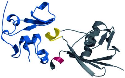
Protective antigen (PA), a component of anthrax toxin, binds receptors on mammalian cells and is activated by a cell surface protease. The resulting active fragment, PA(63), forms ring-shaped heptamers, binds the enzymic moieties of the toxin, and translocates them to the cytosol. Of the four crystallographic domains of PA, domain 1 has been implicated in binding the enzymic moieties; domain 2 is involved in membrane insertion and oligomerization; and domain 4 binds receptor. To determine the function of domain 3, we developed a screen that allowed us to isolate random mutations that cause defects in the activity of PA. We identified several mutations in domain 3 that affect monomer-monomer interactions in the PA(63) heptamer, indicating that this may be the primary function of this domain.


Figures

FIG. 1
FIG. 1
Structure of PA63 with domain 3 mutations. Domain 3 is colored grey. Mutations in the yellow-colored amino acids were isolated in the screen.
FIG. 2
FIG. 2
Native gel electrophoresis of PA mutants. Trypsin-nicked PA (A) or heptameric PA63 (B) was incubated in the presence (+) or absence (−) of LFN and electrophoresed on 4 to 20% acrylamide Tris-glycine gels. The gels were stained with Coomassie blue.
FIG. 3
FIG. 3
Release of 86Rb from CHO-K1 cells by PA and PA mutants. Trypsin-nicked wild-type or mutant PA was incubated with CHO-K1 cells at 4°C. The cells were washed twice with PBS and then exposed to buffer at pH 4.8 to induce channel formation. After 30 min, buffer was removed and its radioactive content was determined by gamma radiation counting. Error bars, standard deviations.
FIG. 4
FIG. 4
Translocation at the cell surface of LFN by wild-type and mutant PA. Trypsin-nicked wild-type or mutant PA was incubated with CHO-K1 cells at 4°C. The cells were washed twice with PBS and incubated with 35S-LFN at 4°C. The cells were exposed to buffer at pH 4.8 and either solubilized with lysis buffer, to determine the amount of bound LFN (− Pronase), or treated with pronase, to determine the amount of translocated LFN (+ Pronase). Radioactive content was measured by scintillation counting. Nonspecific binding of LFN to cells (less than 10%) was subtracted from the experimental measurements to determine specific binding. Error bars, standard deviations.
FIG. 5
FIG. 5
Involvement of domain 3 in the oligomerization of PA63. Domain 1 (amino acids 173 to 258, colored blue) and domain 3 (amino acids 488 to 595 plus amino acids 481 to 487 of domain 2, colored grey) of adjacent monomers are shown. Amino acids 483 to 486 are colored red, and amino acids 510 to 518 are colored yellow.

References

    1. Benson E L, Huynh P D, Finkelstein A, Collier R J. Identification of residues lining the anthrax protective antigen channel. Biochemistry. 1998;37:3941–3948. - PubMed
    1. Blaustein R O, Koehler T M, Collier R J, Finkelstein A. Anthrax toxin: channel-forming activity of protective antigen in planar phospholipid bilayers. Proc Natl Acad Sci USA. 1989;86:2209–2213. - PMC - PubMed
    1. Brossier F, Sirard J, Guidi-Rontani C, Duflot E, Mock M. Functional analysis of the carboxy-terminal domain of Bacillus anthracis protective antigen. Infect Immun. 1999;67:964–967. - PMC - PubMed
    1. Duesbery N S, Webb C P, Leppla S H, Gordon V M, Klimpel K R, Copeland T D, Ahn N G, Oskarsson M K, Fukasawa K, Paull K D, Woude G F V. Proteolytic inactivation of MAP-kinase-kinase by anthrax lethal factor. Science. 1998;280:734–737. - PubMed
    1. Friedlander A M. Macrophages are sensitive to anthrax lethal toxin through an acid-dependent process. J Biol Chem. 1986;261:7123–7126. - PubMed

Publication types

LinkOut - more resources