. 2001 Mar 1;21(5):1444-51.
doi: 10.1523/JNEUROSCI.21-05-01444.2001.

Alpha-1-antichymotrypsin promotes beta-sheet amyloid plaque deposition in a transgenic mouse model of Alzheimer's disease

Affiliations

Alpha-1-antichymotrypsin promotes beta-sheet amyloid plaque deposition in a transgenic mouse model of Alzheimer's disease

L N Nilsson et al. J Neurosci. .

Abstract

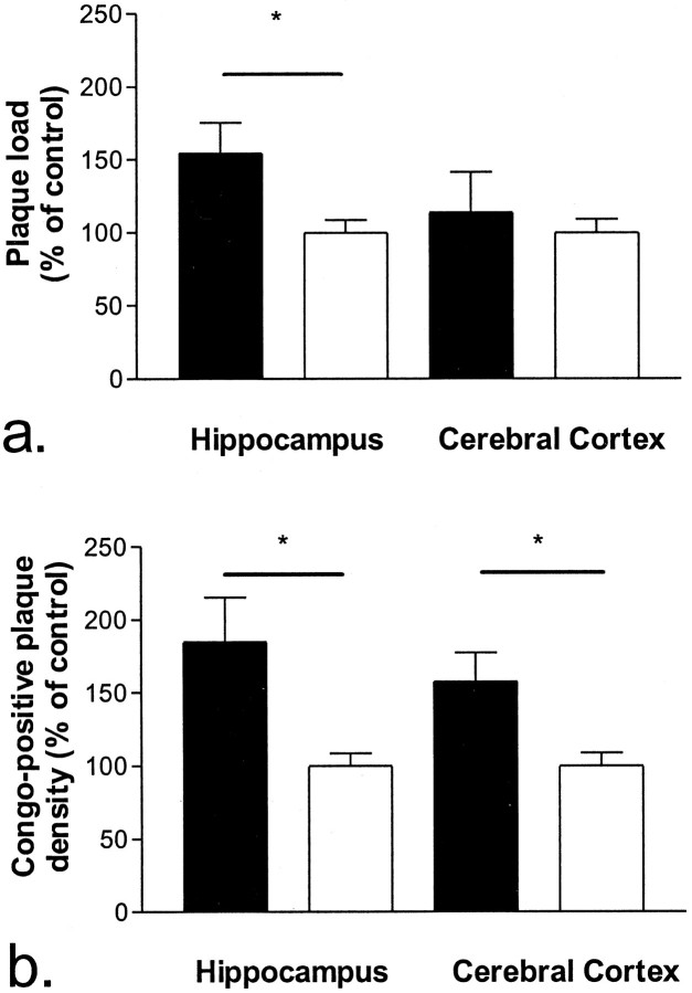
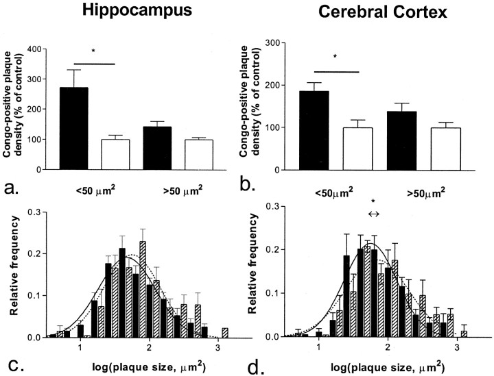
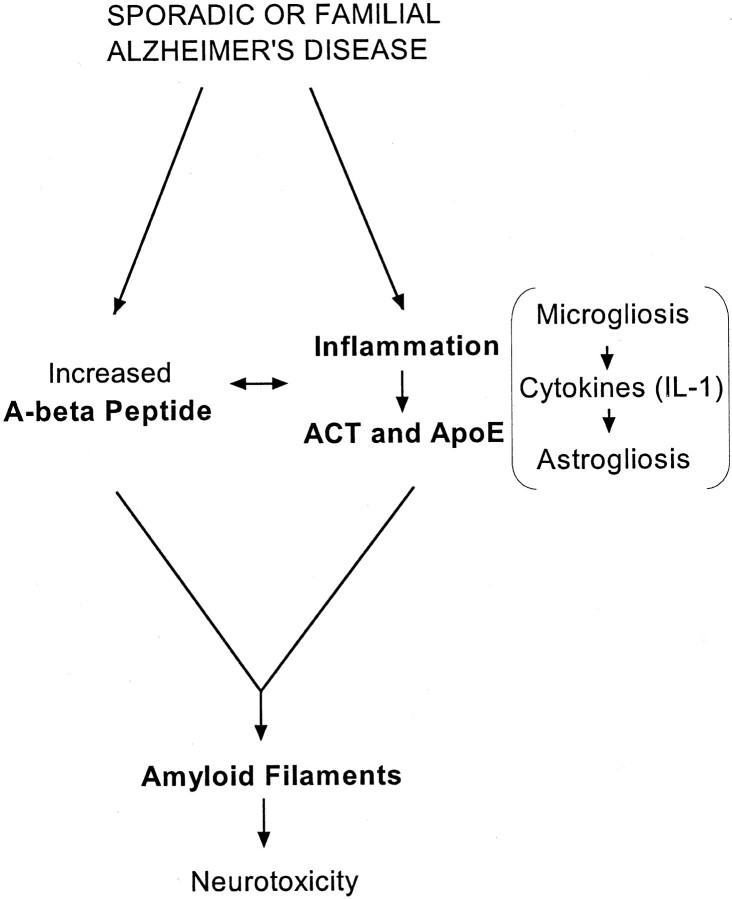
Alpha(1)-antichymotrypsin (ACT), an acute-phase inflammatory protein, is an integral component of the amyloid deposits in Alzheimer's disease (AD) and has been shown to catalyze amyloid beta-peptide polymerization in vitro. We have investigated the impact of ACT on amyloid deposition in vivo by generating transgenic GFAP-ACT-expressing mice and crossing them with the PDGF-hAPP/V717F mice, which deposit amyloid in an age-dependent manner. The number of amyloid deposits measured by Congo Red birefringence was increased in the double ACT/amyloid precursor protein (APP) transgenic mice compared with transgenic mice that only expressed APP, particularly in the hippocampus where ACT expression was highest, and the increase was preceded by elevated total amyloid beta-peptide levels at an early age. Our data demonstrate that ACT promotes amyloid deposition and provide a specific mechanism by which inflammation and the subsequent upregulation of astrocytic ACT expression in AD brain contributes to AD pathogenesis.


Figures

Fig. 1.
Fig. 1.
Expression of ACT mRNA and protein in ACT transgenic mice. a,Immunoprecipitation and Western blot of brain protein extracts from a nontransgenic mouse and ACT-transgenic founder lines (#8782, #8783, and #8784) displaying a protein band (∼68 kDa) that closely comigrates with human serum ACT. b, GFAP/ACT mRNA and GAPDH mRNA expression in brain of a nontransgenic mouse and various tissues of a heterozygous ACT-transgenic mouse showing brain-specific expression of ACT only in the transgenic mouse. These experiments (aand b) were performed on singly ACT transgenic animals without stab wound injury. c, Astrocyte expression and secretion of ACT immunoreactivity was found in the hippocampal formation of heterozygous ACT-transgenic mice (d), whereas both astrocyte staining and the diffuse ACT-immunostaining in the hippocampus seen in ACT transgenic mouse was absent in nontransgenic mouse. e,Colocalization of ACT (brown) and GFAP (blue) immunoreactivity in astrocytes of the stab-wounded ACT(+/−) mice. f, High-power magnification of the tissue section depicted in c showing the morphology of ACT-immunopositive astrocytes. The ACT immunostaining of brain sections from both of these animals was performed 3 d after stab wound injury (c–f). Sections were counterstained with Methyl Green (c, d,f). Scale bars: e, 7.8 μm; f, 12.5 μm.
Fig. 2.
Fig. 2.
Astrocyte-specific expression and plaque association of ACT protein in double APP/ACT transgenic mice.a, Astrogliotic ACT immunostaining along the hippocampal fissure in 6-month-old mouse. b, ACT-immunopositive astrocytes were present in a 10-month-old PDGF-hAPP/V717F(+/−)ACT(+/−) mouse (c) but absent in a 10-month-old PDGF-hAPP/V717F(+/−)ACT(−/−) mouse. d, High-power magnification of ACT-immunopositive astrocytes close to Congo Red-positive amyloid plaques in a hippocampal subregion marked by an arrow in b.e, High-power magnification of an ACT-immunopositive Congo Red-positive amyloid plaque in the hippocampus of a PDGF-hAPP/V717F(+/−) ACT(+/−) mouse at 10 months of age (f) displaying birefringence under polarized light. Scale bars: a, e,f, 7.8 μm; d, 12.5 μm.
Fig. 3.
Fig. 3.
Increased total Congo Red-positive amyloid plaque load (a) and numerical plaque density (b) in the hippocampus and the cerebral cortex of 10-month-old PDGF-hAPP/V717F(+/−)ACT(+/−) mice (n= 7; solid bar), compared with age-matched PDGF-hAPP/V717F (+/−)ACT(−/−) mice (n = 6;open bar). The results are expressed as percentage of the control group (the PDGF-hAPP/V717F (+/−)ACT(−/−) genotype mice) and represent mean ± SEM.
Fig. 4.
Fig. 4.
Plaque density analysis. Congo-positive amyloid plaques in the hippocampus (a,c) and the cerebral cortex (b,d) were stratified according to their sizes. There was an increased numerical density of small plaques (<50 μm2) in the 10-month-old PDGF-hAPP/V717F(+/−) ACT(+/−) mice (n = 7;solid bar) compared with the age-matched PDGF-hAPP/V717F(+/−) ACT(−/−) mice (n = 6;open bar) in the hippocampus (a) and cerebral cortex (b). Shown below bar graphs are the corresponding relative frequency histograms of plaque size distribution in mice of the PDGF-hAPP/V717F(+/−) ACT(+/−) (solid bar) and the PDGF-hAPP/V717F(+/−), ACT(−/−) genotypes (hatched bar) in the hippocampus (c) and the cerebral cortex (d). Superimposed is the best fit of our data to a Gaussian distribution for mice of the PDGF-hAPP/V717F(+/−)ACT(+/−) (solid line) and the PDGF-hAPP/V717F(+/−) ACT(−/−) genotypes (broken line). The center of the Gaussian distribution curve for the PDGF-hAPP/V717F(+/−)ACT(+/−) mice was shifted toward a smaller average plaque size in the hippocampus (c) as well as the cerebral cortex (d). The results displayed are expressed as percentage of the control group [the PDGF-hAPP/V717F(+/−)ACT (−/−)] genotype and represent mean ± SEM.
Fig. 5.
Fig. 5.
Increased total amyloid β-peptide (Aβ) and Aβ1–42 levels in the hippocampus of 3-month-old PDGF-hAPP/V717F(+/−)ACT(+/−) mice (n = 10;solid bar) compared with age-matched PDGF hAPP/V717F(+/−)ACT(−/−) mice (n = 14;open bar).
Fig. 6.
Fig. 6.
Pathogenic pathway leading to Alzheimer's disease.

References

    1. Abraham CR, Selkoe DJ, Pottter H. Immunohistochemical identification of the serine protease inhibitor α1-antichymotrypsin in the brain amyloid deposits of Alzheimer's disease. Cell. 1988;52:487–501. - PubMed
    1. Abraham CR, Shirahama T, Potter H. α1-antichymotrypsin is associated solely with amyloid deposits containing the β-protein. Amyloid and cell localization of α1-antichymotrypsin. Neurobiol Aging. 1990;11:123–129. - PubMed
    1. Akiyama H, Barger S, Barnum S, Bradt B, Bauer J, Cole GM, Cooper NR, Eikelenboom P, Emmerling M, Fiebich BL, Finch CE, Frautschy S, Griffin WS, Hampel H, Hull M, Landreth G, Lue L, Mrak R, Mackenzie IR, McGeer PL, O'Banion MK, Pachter J, Pasinetti G, Plata-Salaman C, Rogers J, Rydel R, Shen Y, Streit W, Strohmeyer R, Tooyoma I, Van Muiswinkel FL, Veerhuis R, Walker D, Webster S, Wegrzyniak B, Wenk G, Wyss-Coray T. Inflammation and Alzheimer's disease. Neurobiol Aging. 2000;21:383–421. - PMC - PubMed
    1. Bales KR, Verina T, Dodel RC, Du Y, Altstiel L, Bender M, Hyslop P, Johnstone EM, Little SP, Cummins DJ, Piccardo P, Ghetti B, Paul SM. Lack of apolipoprotein E dramatically reduces amyloid β-peptide deposition. Nat Genet. 1997;17:263–264. - PubMed
    1. Bales KR, Verina T, Cummins DJ, Du Y, Dodel RC, Saura J, Fishman CE, Delong CA, Piccardo P, Petegnief V, Ghetti B, Paul SM. Apolipoprotein E is essential for amyloid deposition in the APPV717F transgenic mouse model of Alzheimer's disease. Proc Natl Acad Sci USA. 1999;96:15233–15238. - PMC - PubMed

Publication types

MeSH terms

Substances