. 2001 Mar 1;21(5):1452-63.
doi: 10.1523/JNEUROSCI.21-05-01452.2001.

Molecular and physiological diversity of nicotinic acetylcholine receptors in the midbrain dopaminergic nuclei

Affiliations

Molecular and physiological diversity of nicotinic acetylcholine receptors in the midbrain dopaminergic nuclei

R Klink et al. J Neurosci. .

Abstract

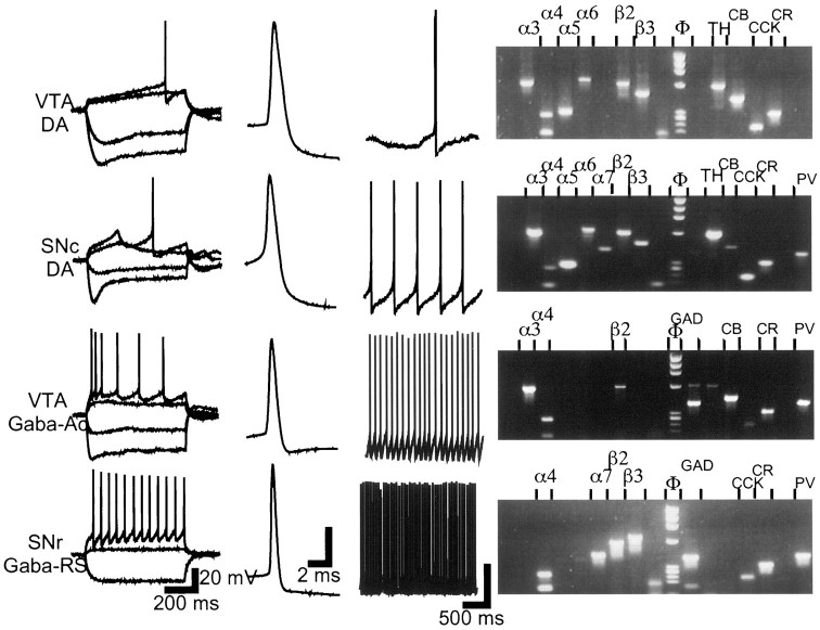
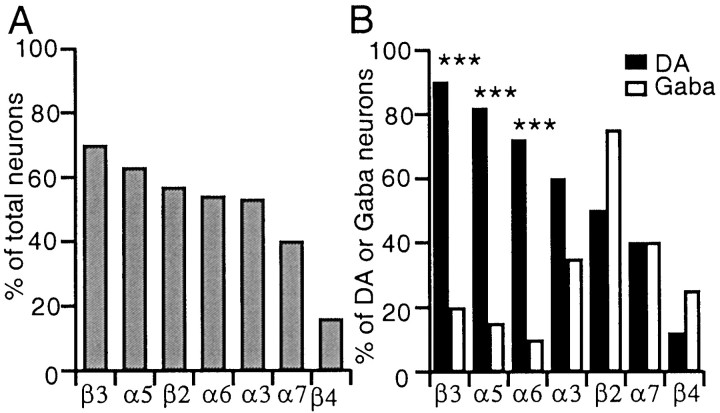
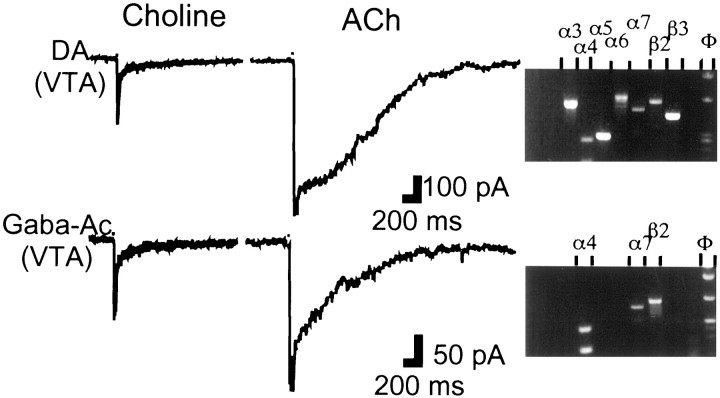
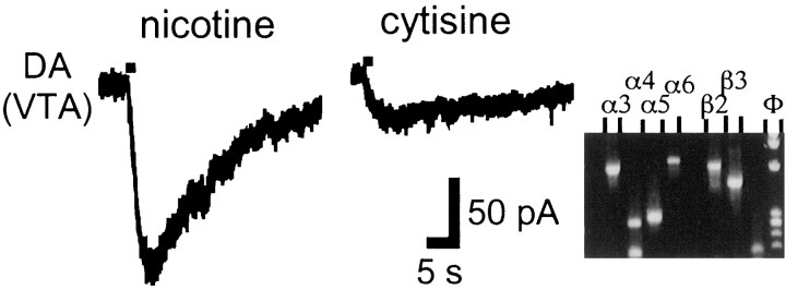
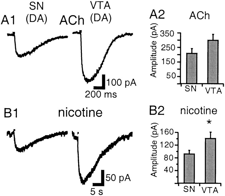
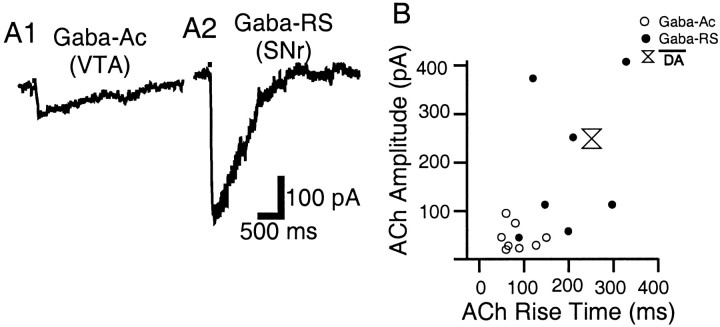
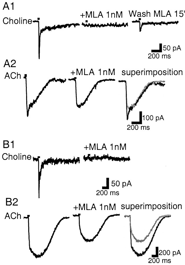
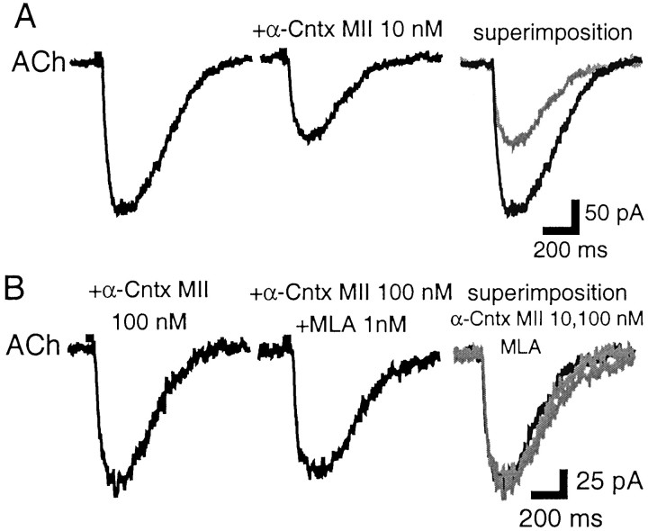
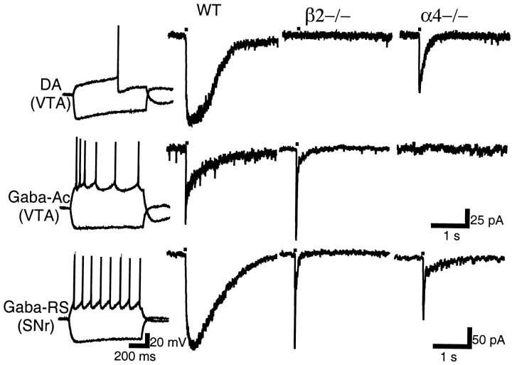
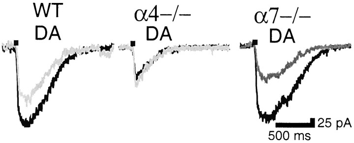
Nicotinic acetylcholine receptors (nAChRs) on dopaminergic (DA) and GABAergic (Gaba) projection neurons of the substantia nigra (SN) and ventral tegmental area (VTA) are characterized by single-cell RT-PCR and patch-clamp recordings in slices of rat and wild-type, beta2-/-, alpha4-/-, and alpha7-/- mice. The eight nAChR subunits expressed in these nuclei, alpha3-7 and beta2-4, contribute to four different types of nAChR-mediated currents. Most DA neurons in the SN and VTA express two nAChR subtypes. One is inhibited by dihydro-beta-erythroidine (2 microm), alpha-conotoxin MII (10 nm), and methyllycaconitine (1 nm) but does not contain the alpha7 subunit; it possesses a putative alpha4alpha6alpha5(beta2)(2) composition. The other subtype is inhibited by dihydro-beta-erythroidine (2 microm) and has a putative alpha4alpha5(beta2)(2) composition. Gaba neurons in the VTA exhibit a third subtype with a putative (alpha4)(2)(beta2)(3) composition, whereas Gaba neurons in the SN have either the putative (alpha4)(2)(beta2)(3) oligomer or the putative alpha4alpha6alpha5(beta2)(2) oligomer. The fourth subtype, a putative (alpha7)(5) homomer, is encountered in less than half of DA and Gaba neurons, in the SN as well as in the VTA. Neurons in the DA nuclei thus exhibit a diversity of nAChRs that might differentially modulate reinforcement and motor behavior.


Figures

Fig. 1.
Fig. 1.
Electrophysiological and molecular profile of VTA and SN neurons. Electrophysiological classes were distinguished on the basis of intrinsic membrane potential and firing properties in response to current steps applied from −64 mV (first panel from left); on action potential waveform and duration, measured at rheobase current (second panel); and on firing frequency at the resting membrane potential for neurons that were spontaneously active (third panel). DA neurons in the SNc and SNr were indistinguishable; they exhibited strong Ihactivation and slow potential oscillations in the subthreshold range. DA neurons in the VTA showed a less pronouncedIh activation and a slow ramp potential before firing initiation. Gaba-Ac neurons in the VTA were characterized by marked spike-frequency accommodation in response to depolarizing current steps. Gaba-RS neurons in the SNr fired with a regular discharge pattern, at all amplitudes of depolarizing current steps and from depolarized as well as hyperpolarized holding potentials. Vertical calibration bar is 20 mV in all three panels. The right panel shows agarose gels of the PCR amplification products corresponding to the illustrated neuron. Only detected products are labeled (left to right): nicotinic subunits α2–7 and β2–4, marker, GAD (GAD 65 and GAD 67), TH, CB, CCK, CR, NT, and PV. The gel for the GABA-Ac neuron (third from top) shows a faint TH band; such (rare) TH bands were discounted. When GAD and TH were coexpressed (see Results) both bands were of equal intensity.
Fig. 2.
Fig. 2.
Frequency distribution of mRNAs for neurotransmitter synthesizing enzymes, neuropeptides, and Ca2+-binding proteins in SN and VTA.A, Percentage of neurons coexpressing GAD and TH mRNA versus age; numbers at the base of histogram bars indicate the total number of neurons in each age group bin (2 d).B1, Percentage of neurons expressing the neuropeptides CCK and NT and the Ca2+-binding proteins CR, PV, and CB. B2, Percentage of DA and Gaba neurons that are CCK positive. CCK is preferentially expressed in DA neurons.B3, Percentage of CB-positive neurons in the SNc, SNr, and VTA. CB was found preferentially in the VTA (**p < 0.01).
Fig. 3.
Fig. 3.
Frequency distribution of nicotinic subunit mRNAs in the SN and VTA. A, Nicotinic subunits are sorted by order of decreasing prevalence: subunit α4 was present in all neurons, and subunit α2 was present in none (data not shown).B, The same distribution segregated with respect to neuron class. β3, α5, and α6 mRNAs are significantly more prevalent in DA than in Gaba neurons (***p < 0.001).
Fig. 4.
Fig. 4.
Neurons containing subunit α7 mRNA exhibit fast currents gated by choline and ACh. Choline (10 mm; 30 msec) elicits identical current waveforms in DA and Gaba neurons (left panel). In the same neurons, ACh (1 mm; 30 msec) elicits different current waveforms (middle panel), in both of which a fast component can be recognized. In this and subsequent figures, currents were evoked from a holding potential of −70 mV, and the width of the black box at the beginning of each trace indicates duration of pressure-pulse application. Agarose gels (right panel) corresponding to the DA and Gaba neuron show the presence of subunit α7 mRNA (gels truncated after the nAChR subunit wells).
Fig. 5.
Fig. 5.
Nicotine elicits larger currents than cytisine in neurons lacking subunit β4. Nicotine (20 μm;left panel) and cytisine (20 μm;middle panel) were pressure-applied for 1 sec. Agarose gel (right panel; truncated after the nAChR subunit wells) shows the absence of subunit β4.
Fig. 6.
Fig. 6.
Nicotinic currents in DA neurons of the SN and VTA. A1, ACh (1 mm; 30 msec) elicits currents of similar waveform in the VTA and SN, characterized by a round peak and sigmoid decay. B1, In the same neurons, nicotine (20 μm; 300 msec) elicits much longer lasting currents. A2, Mean ACh-elicited current amplitude in the SN and VTA. B2, Mean nicotine-elicited current amplitude in the SN and VTA. Nicotine induces larger currents in the VTA than in the SN (*p < 0.05).
Fig. 7.
Fig. 7.
ACh-elicited currents in Gaba neurons of the SN and VTA. A1, In the Gaba-Ac subclass of the VTA, ACh elicits current waveforms characterized by a sharp peak, exponential decay, and small amplitude. A2, In the Gaba-RS subclass of the SNr, the ACh current waveform is often slower and of larger amplitude. B, ACh elicited current amplitude plotted versus corresponding rise time in Gaba-Ac (○) and Gaba-RS neurons (●); mean ACh-elicited current amplitude versus mean rise time in DA neurons was also reported (double triangle).
Fig. 8.
Fig. 8.
MLA affects two different types of currents.A1, In a VTA DA neuron, MLA (1 nm) blocks the choline-gated current, which partially recovers after a 15 min wash period. A2, In the same neuron, MLA blocks the fast component of the ACh-gated current without affecting the slower component. B1, In another VTA DA neuron, MLA (1 nm) blocks the choline-gated current. B2, In the same neuron, the ACh-gated current does not show a clear fast component; however, MLA inhibits the slow peak current occurring later. In the superimposition panels, MLA trace is ingray.
Fig. 9.
Fig. 9.
α-conotoxin MII inhibits the MLA-sensitive component of ACh-gated currents. A, In an SNc DA neuron, α-conotoxin MII (10 nm) inhibits the ACh-gated current. In the panel labeled superimposition, the MLA trace is in gray. B, In the same neuron, increased concentration of α-conotoxin MII (100 nm) does not result in greater inhibition (note change in scale); addition of MLA (1 nm) to the α-conotoxin MII (100 nm) also has no effect (middle). In thesuperimposition panel, the α-conotoxin MII (100 nm) and MLA traces are in gray. α-Cntx MII, α-Conotoxin MII.
Fig. 10.
Fig. 10.
Neuron classes and their ACh-elicited currents in the SN and VTA of WT and nAChR subunit null mutant mice. Electrophysiological classes (left panel) and ACh-elicited current waveforms in WT (second panel fromleft) were similar to those described in rat. In β2−/− mice, only a fast, α7-homomeric type of ACh-gated current could be recorded in the illustrated neuronal subclasses (third panel). In α4−/− mice, a slow current could be elicited only in DA neurons (right panel). Calibration in second row applies also to top row.
Fig. 11.
Fig. 11.
MLA-sensitive currents in WT and nAChR subunit null mutant mice. Left, DA neurons in WT mice are partially inhibited by MLA (1 nm); middle, MLA sensitivity is abolished in α4−/− mice; right, MLA sensitivity is not affected in α7−/− mice. MLA traces are ingray.

References

    1. Alkondon M, Albuquerque EX. Diversity of nicotinic acetylcholine receptors in rat hippocampal neurons. I. Pharmacological and functional evidence for distinct structural subtypes. J Pharmacol Exp Ther. 1993;265:1455–1473. - PubMed
    1. Alkondon M, Pereira EF, Cortes WS, Maelicke A, Albuquerque EX. Choline is a selective agonist of α7 nicotinic acetylcholine receptors in the rat brain neurons. Eur J Neurosci. 1997;9:2734–2742. - PubMed
    1. Arroyo-Jiménez MM. PhD thesis. Universidad Miguel Hernández, Albacete, Spain; 2000. Localization of the α4 and β3 subunits of the neuronal nicotinic acetylcholine receptor in the rodent central nervous system.
    1. Baron JA. Cigarette smoking and Parkinson's disease. Neurology. 1986;36:1490–1496. - PubMed
    1. Berke JD, Hyman SE. Addiction, dopamine, and the molecular mechanisms of memory. Neuron. 2000;25:515–532. - PubMed

Publication types

MeSH terms

Grants and funding