. 2001 Mar 1;21(5):1523-31.
doi: 10.1523/JNEUROSCI.21-05-01523.2001.

Altered electrical properties in Drosophila neurons developing without synaptic transmission

Affiliations

Altered electrical properties in Drosophila neurons developing without synaptic transmission

R A Baines et al. J Neurosci. .

Abstract

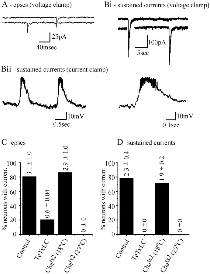
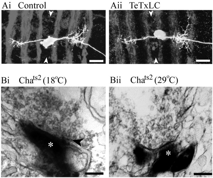
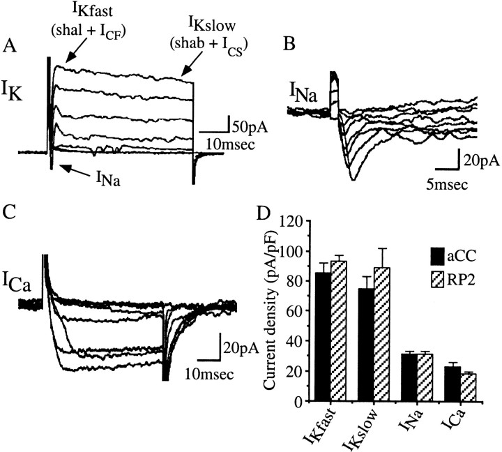
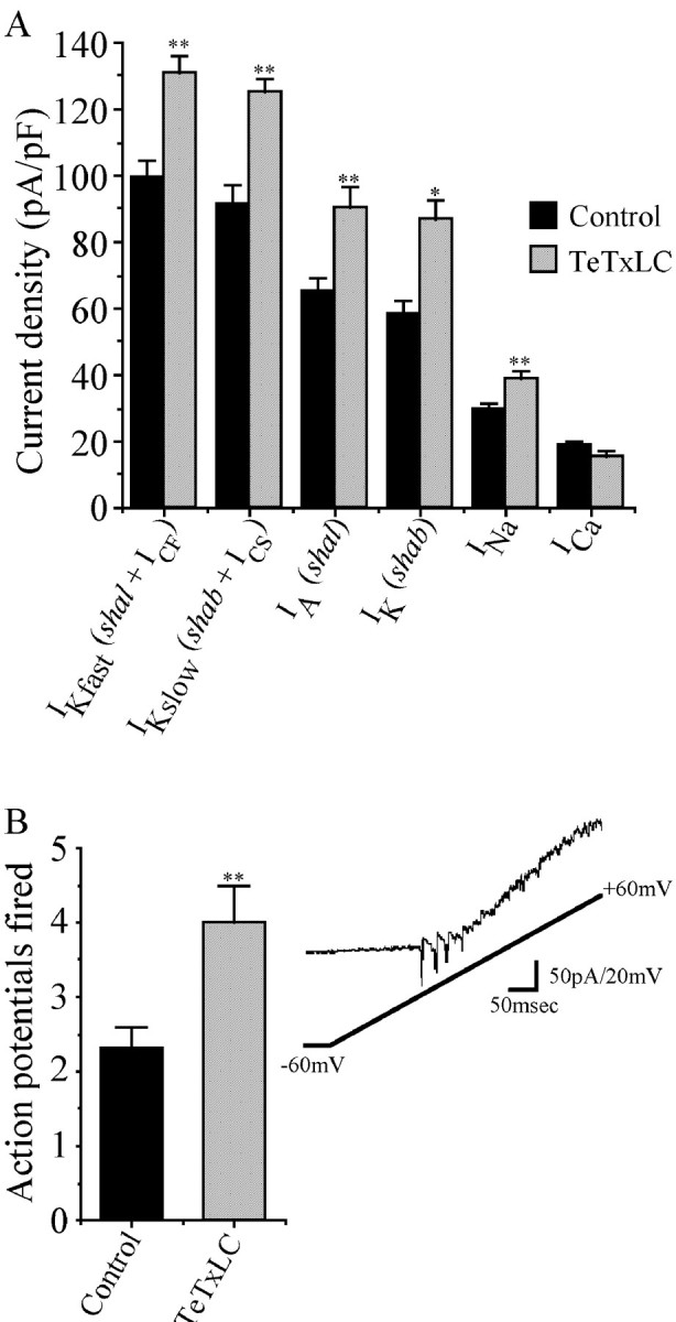
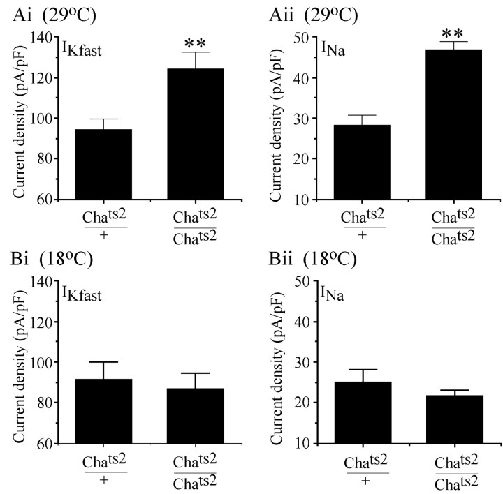
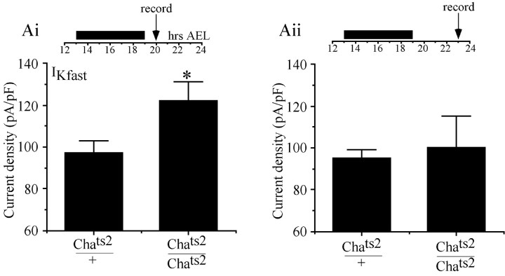
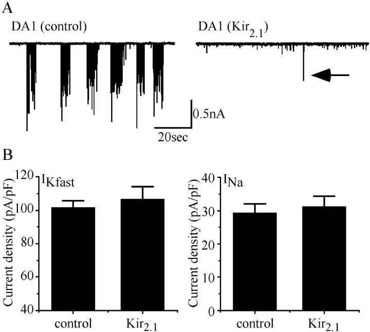
We examine the role of synaptic activity in the development of identified Drosophila embryonic motorneurons. Synaptic activity was blocked by both pan-neuronal expression of tetanus toxin light chain (TeTxLC) and by reduction of acetylcholine (ACh) using a temperature-sensitive allele of choline acetyltransferase (Cha(ts2)). In the absence of synaptic activity, aCC and RP2 motorneurons develop with an apparently normal morphology and retain their capacity to form synapses. However, blockade of synaptic transmission results in significant changes in the electrical phenotype of these neurons. Specifically, increases are seen in both voltage-gated inward Na(+) and voltage-gated outward K(+) currents. Voltage-gated Ca(2+) currents do not change. The changes in conductances appear to promote neuron excitability. In the absence of synaptic activity, the number of action potentials fired by a depolarizing ramp (-60 to +60 mV) is increased and, in addition, the amplitude of the initial action potential fired is also significantly larger. Silencing synaptic input to just aCC, without affecting inputs to other neurons, demonstrates that the capability to respond to changing levels of synaptic excitation is intrinsic to these neurons. The alteration to electrical properties are not permanent, being reversed by restoration of normal synaptic function. Whereas our data suggest that synaptic activity makes little or no contribution to the initial formation of embryonic neural circuits, the electrical development of neurons that constitute these circuits seems to depend on a process that requires synaptic activity.


Figures

Fig. 1.
Fig. 1.
Synaptic input to aCC and RP2 is identical. Passive recordings from wild-type aCC and RP2 neurons voltage clamped at −60 mV show the presence of two types of synaptic input.A, Discrete inward currents of small amplitude (18 pA average) and short duration (20–40 msec) are visible in the majority of recordings. These events have previously been shown to fulfill the criteria expected of EPSCs (Baines and Bate, 1998). Bi, Recordings from aCC/RP2 also reveal the presence of relatively slow- and large-amplitude sustained inward currents. Bii, In current-clamp mode, sustained currents produce large depolarizations that trigger the firing of action potentials in aCC/RP2. Action potentials are clearly visible at a slower time base (right panel). C and D show the proportion of aCC/RP2 neurons that exhibit EPSCs (C) and sustained inward currents (D) in the various genetic backgrounds used in this study; control (including WT, scabrous GAL4, and UAS-TeTxLC), TeTxLC expression, and Chats2 at either 18°C (permissive temperature) or 29°C (restrictive temperature). Bars represent the percentage of neurons in which these respective currents were observed, whereas values given represent the average frequency (per minute) observed in each case (n = 27, 10, 7, and 7 neurons, respectively). Frequency of EPSCs and sustained currents in TeTxLC andChats2 (29°C) are significantly different from control and Chats2(18°C), respectively (p ≤ 0.01).
Fig. 2.
Fig. 2.
Morphology of aCC and presynaptic terminals develop independently of synaptic activity. A, Confocal projections of aCC neurons retrogradely labeled by DiI in both control (inactive TeTxLC; Ai) and when evoked synaptic activity is blocked (active TeTxLC; Aii). No differences in morphology are attributable to the absence of synaptic activity (Table1). Neurons were labeled at 20–21 hr AEL, and preparations have been counterstained with antibody to FasII to visualize the axon scaffold.Arrowheads indicate the position of the midline. Scale bar, 10 μm; anterior is topmost. B, Electron micrographs of photoconverted DiI-labeled aCC neurons show labeled profiles (asterisks) in control (Chats2 at 18°C; Bi) and in CNS in which evoked synaptic activity is absent (Chats2 at 29°C;Bii). Sites of synaptic input to aCC were identified by an accumulation of clear synaptic vesicles, some of which are docked to the presynaptic membrane, immediately adjacent to such labeled profiles. At this stage of development, synaptic elements such as T-bars are rare (Bi, arrow), although an increased electron density of the presynaptic membrane is often visible (Bii). Scale bar, 200 nm.
Fig. 3.
Fig. 3.
Voltage-dependent ion channel characteristics in aCC and RP2. A, Whole-cell voltage clamp of aCC/RP2 reveals the presence of at least two voltage-activated outward K+ macro currents (IKfastand IKslow) and a voltage-activated inward sodium current (INa). These neurons also exhibit voltage-activated inward calcium currents that are masked by IK under these conditions.IKfast and IKsloware composed of at least four individual currents, shal+ ICF and shab +ICS, respectively, see Results for details. B, Blocking both outwardIK and inward ICaisolates the voltage-activated inward INa.C, Isolation of voltage-activatedICa was achieved by blocking voltage-activated IK andINa currents. ICawas measured using barium as the permeant ion (see Baines and Bate, 1998 for discussion of use of this ion). Currents shown are from aCC in embryos at ∼20 hr AEL. Currents were evoked using voltage steps (15 mV increments; range, −60 to +45 mV; 50 msec) applied from a conditioning prepulse of −90 mV (100 msec duration). Traces shown are the average of five trials. D, Peak current density, normalized to membrane capacitance, for the currents isolated in aCC and RP2. No currents are significantly different between aCC and RP2. Values shown are mean ± SE; n ≥ 8.
Fig. 4.
Fig. 4.
Absence of synaptic input changes aCC/RP2 electrical properties. A, Peak current density for the voltage-activated ion currents isolated in aCC/RP2 in either a TeTxLC-expressing CNS (no synaptic activity) or a control (inactive TeTxLC) CNS in which synaptic activity is normal. Currents have been normalized to membrane capacitance. The absence of synaptic activity results in a significant increase in IKfast(shal + ICF),IKslow (shab +ICS), IA(shal), IK(shab), and INa(para), but not ICa. Values are mean ± SE; n > 8; *p ≤ 0.05; **p ≤ 0.01.B, A ramp depolarization, generated using voltage clamp, fires significantly more action potentials in aCC/RP2 neurons that have been deprived of synaptic input during their development (TeTxLC) compared with controls (inactive TeTxLC). Values are mean ± SE;n = 11; p ≤ 0.01.Inset shows a typical current recording from an aCC neuron in a CNS lacking synaptic activity; four action potentials are fired, which become progressively smaller because of the increasing membrane potential.
Fig. 5.
Fig. 5.
Selective blockade of synaptic input mimics TeTxLC expression. A,Chats2embryos raised at 29°C result in a paralytic phenotype caused by a significant reduction of ACh. At this temperature, aCC/RP2 show no synaptic input (Fig. 1C,D), although their capability to release neurotransmitter is not affected. Under these conditions, aCC/RP2 show increased current densities of bothIKfast (shal +ICF) (Ai) andINa (Aii) compared with controls (Chats2/+). Flies were allowed to lay eggs at 18°C for 2 hr periods after which embryos were raised at 29°C. B, At a continuous developmental temperature of 18°C, Chats2 embryos are viable and recordings show that aCC/RP2 receive normal levels of synaptic input (Fig. 1C,D). In addition,IKfast (Bi) andINa (Bii) are not increased above controls (Chats2/+). Values shown represent mean ± SE; n ≥ 8. Values forIKfast and INa at 29°C are significantly different from controls (p ≤ 0.01).
Fig. 6.
Fig. 6.
Alterations in electrical properties produced by the removal of synaptic input are reversible. Ai, By shifting Chats2 embryos from 18°C (permissive temperature) to 29°C (restrictive temperature,black bar) between 13 and 19 hr AEL, it is possible to suppress synaptic input to aCC/RP2 during the period in which electrogenesis occurs in these neurons. After this treatment embryos were returned to 18°C. Recordings at 20 hr AEL show that peakIKfast (shal +ICF) density is significantly increased (p ≤ 0.05). Aii,This temperature shift, however, also delays hatching until ∼23–24 hr AEL. Recordings from aCC/RP2 in embryos on hatching show that peakIKfast density has decreased to control levels. Values shown represent mean ± SE; n≥ 8. All timings shown are normalized to development at 25°C (21 hr development time, 41 hr at 18°C).
Fig. 7.
Fig. 7.
Suppression of action potentials does not influence electrical properties. A, Expression of Kir2.1 in aCC using RRC-GAL4 blocks the ability of this neuron to fire action potentials as evidenced by the significant reduction of EJCs recorded in its target muscle (DA1) compared with controls (UAS parental line shown). In the trace shown (right), just one EJC is visible (arrow), although smaller events caused by spontaneous release of neurotransmitter are unaffected. Frequency of synaptic input to aCC is unaffected by expression of Kir2.1 (see Results), although sustained currents fail to trigger action potentials.B, Measurement of peak IKfast(shal + ICF) andINa in aCC in either control (Gal4 and UAS parental lines) or when expressing Kir2.1 shows no significant differences. Values shown represent mean ± SE;n ≥ 8.

References

    1. Baines RA, Bate M. Electrophysiological development of central neurons in the Drosophila embryo. J Neurosci. 1998;18:4673–4683. - PMC - PubMed
    1. Baines RA, Robinson SG, Fujioka M, Jaynes JB, Bate M. Postsynaptic expression of tetanus toxin light chain blocks synaptogenesis in Drosophila. Curr Biol. 1999;9:1267–1270. - PMC - PubMed
    1. Bate M. Development of motor behaviour. Curr Opin Neurobiol. 1999;9:670–675. - PubMed
    1. Bito H. The role of calcium in activity-dependent neuronal gene expression. Cell Calcium. 1998;23:143–150. - PubMed
    1. Brand AH, Perrimon N. Targeted gene expression as a means of altering cell fates and generating dominant phenotypes. Development. 1993;118:401–415. - PubMed

Publication types

MeSH terms

LinkOut - more resources