. 2001 Mar 1;21(5):1767-78.
doi: 10.1523/JNEUROSCI.21-05-01767.2001.

Extrinsic modulation and motor pattern generation in a feeding network: a cellular study

Affiliations

Extrinsic modulation and motor pattern generation in a feeding network: a cellular study

V A Straub et al. J Neurosci. .

Abstract

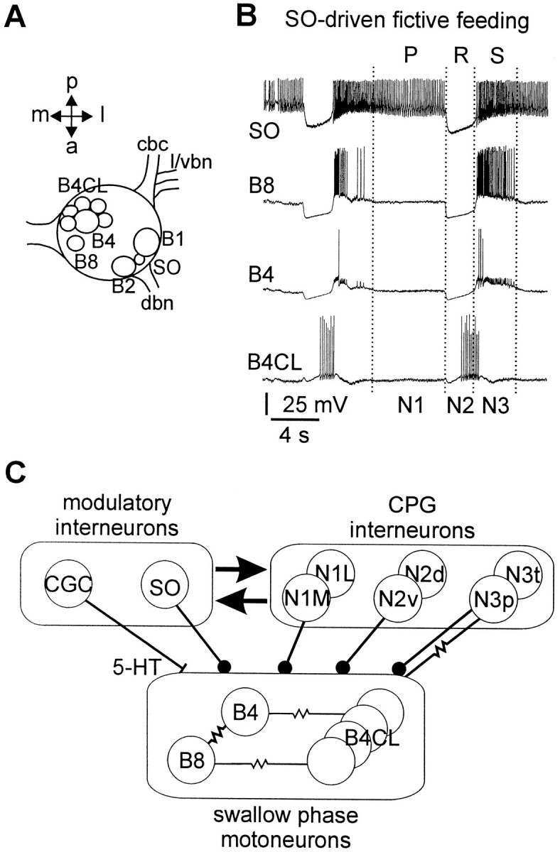
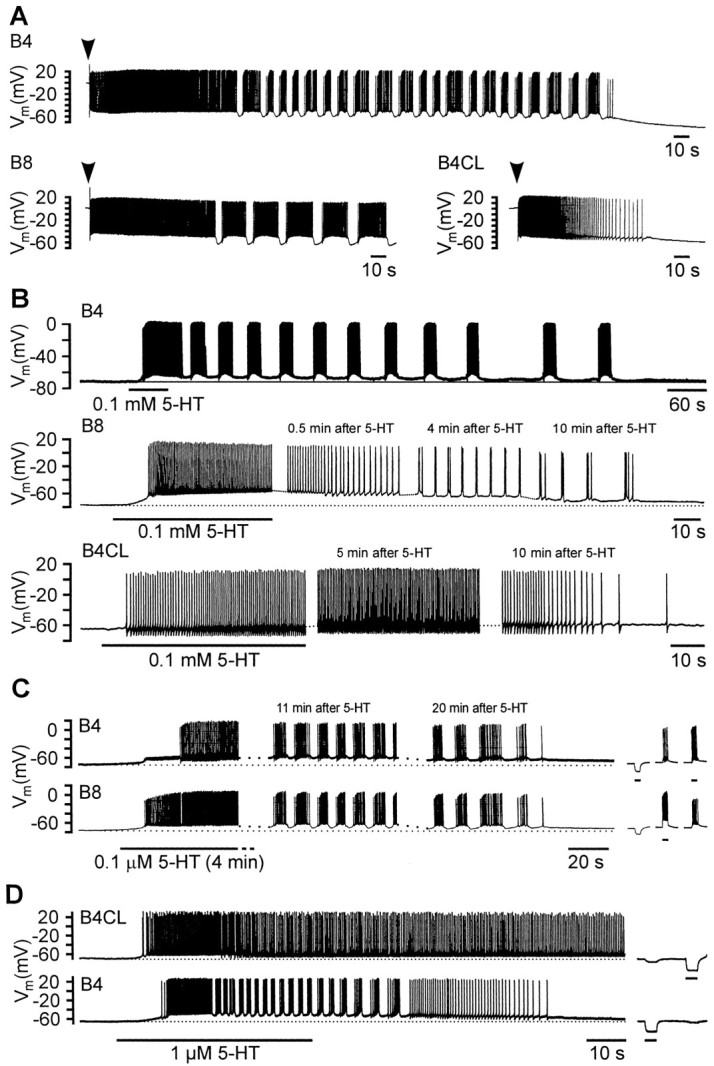
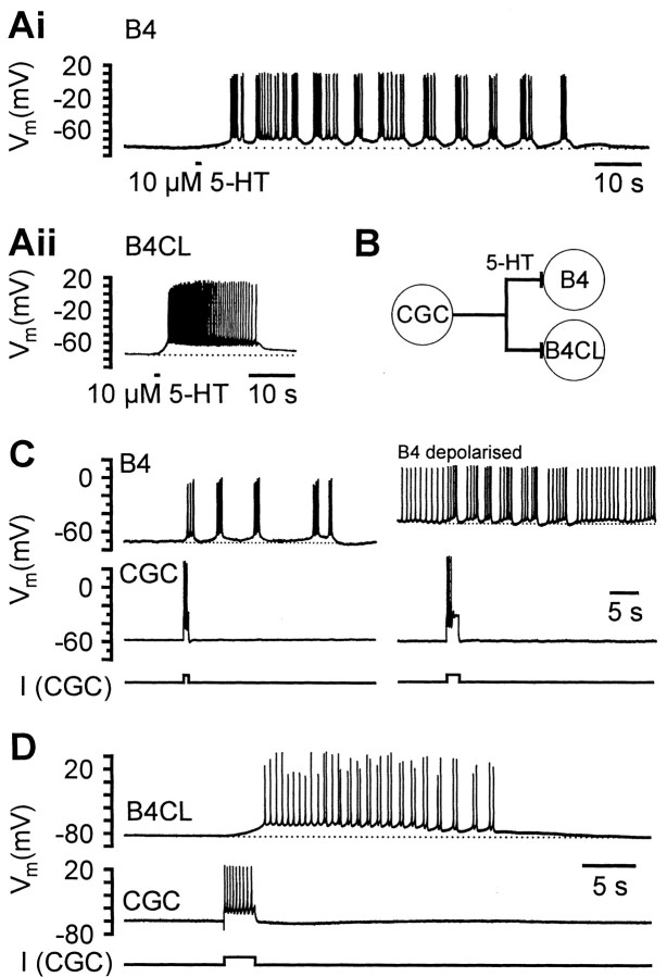
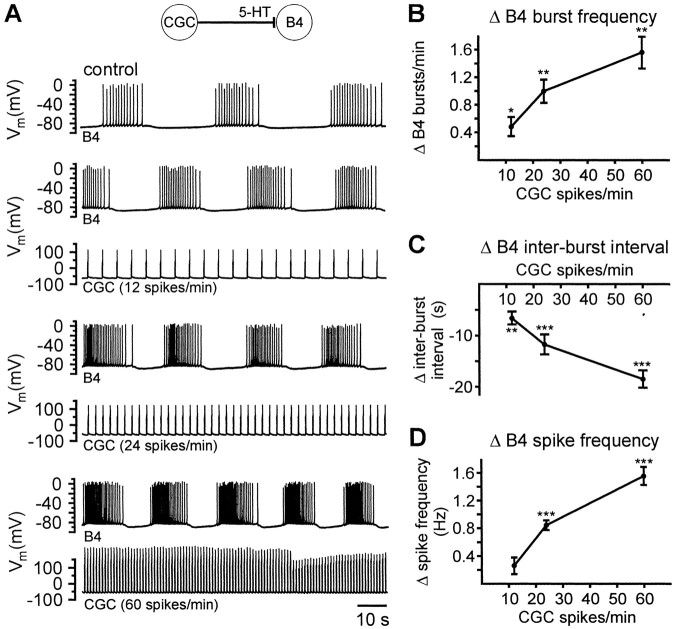
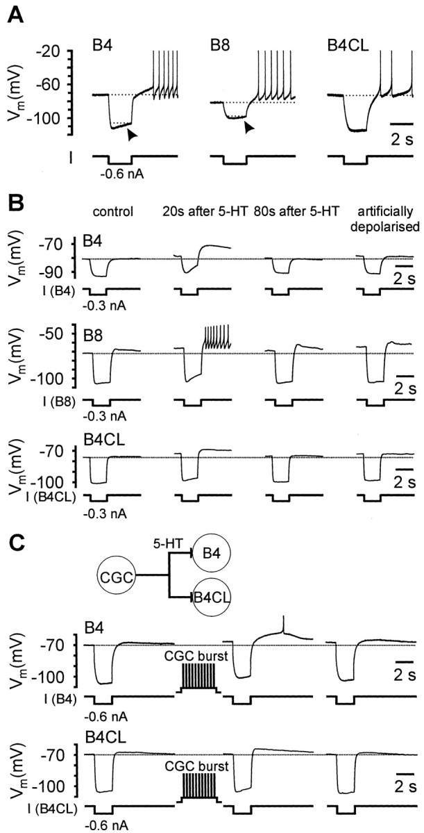
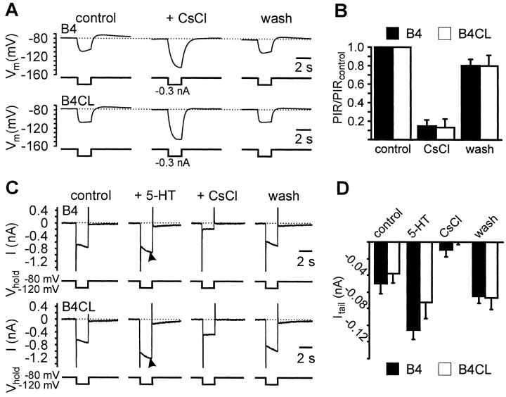
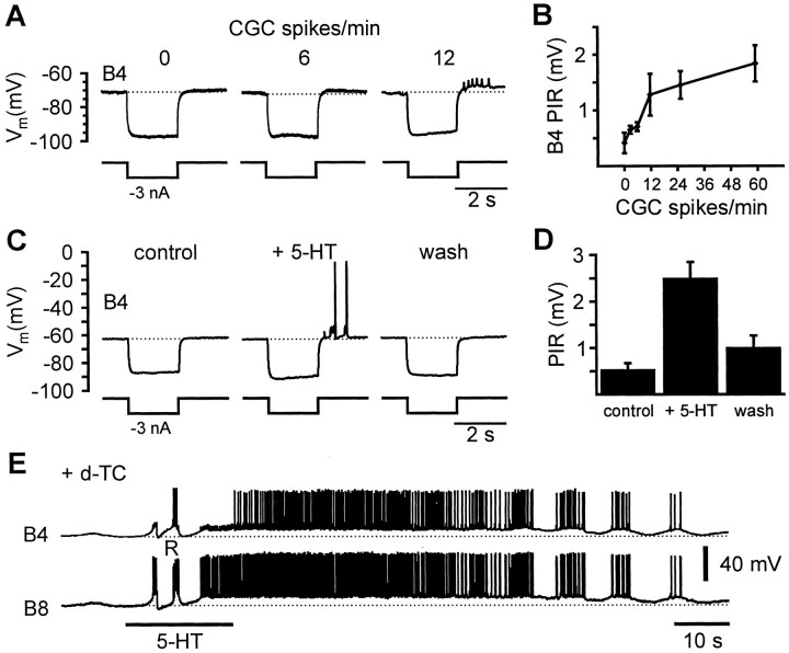
Systems level studies have shown that the paired serotonergic cerebral giant cells (CGCs) of gastropod mollusks have important extrinsic modulatory actions on the central pattern generator (CPG) underlying rhythmic ingestion movements. Here we present the first study that investigates the modulatory actions of the CGCs and their released transmitter 5-HT on the CPG at the cellular level. In the snail, Lymnaea, motoneurons such as the B4, B8, and B4CL cells are part of the feeding CPG and receive serotonergic synaptic inputs from CGCs. These motoneurons were used to investigate the effect of serotonergic modulation on endogenous cellular properties of CPG neurons. Cells were isolated from the intact nervous system, and their properties were examined by pharmacological methods in cell culture. Motoneurons were also grown in coculture with CGCs to compare 5-HT effects with CGC stimulation. Three distinct modulatory effects of exogenously applied 5-HT/CGC activity were seen: all three motoneuron types were depolarized by 5-HT for prolonged periods leading to firing. Conditional bursting accompanied this depolarization in the B4/B8 cells, but not in B4CL cells. The frequency of the bursting was increased with increased CGC tonic firing. An increase in the size of postinhibitory rebound (PIR) occurred with 5-HT application in all three cell types, because of an increase in a CsCl-sensitive, hyperpolarization-activated inward current. Similar modulatory effects on membrane potential, endogenous bursting, and PIR properties could be observed in the intact nervous system and were necessary for motoneuron activation during feeding. Part of the systems gating and frequency control functions of the CGCs appear to be caused by these modulatory effects on feeding motoneurons.


Figures

Fig. 1.
Fig. 1.
Location, firing patterns, and synaptic connectivity of retraction phase neurons in the buccal ganglia ofLymnaea. A, Schematic dorsal view of the left buccal ganglion showing the soma positions of B4, B8, and B4CL retraction phase neurons. The position of the B1 and B2 neurons is shown for reference purposes. The slow oscillator (SO) can be located either in the left or right buccal ganglion between the B1 and B2 neuron. cbc, Cerebrobuccal connective;l/vbn, lateral/ventral buccal nerve; dbn,dorsal buccal nerve; a, anterior; p,posterior; l, lateral; m, medial.B, Simultaneous recording from retraction phase motoneurons B4CL, B4, and B8 in the isolated nervous system showing two cycles of fictive feeding. The fictive feeding pattern was driven by injection of a constant depolarizing current into the SO (top trace). The three phases of the feeding pattern (P/N1 protraction, R/N2 rasp, and S/N3 swallow) are marked above and below the traces. C, Schematic diagram of the known connections between retraction phase motoneurons, feeding CPG interneurons, and modulatory interneurons. Circlesindicate inhibitory synapses, bars indicate excitatory synapses, and resistor symbols indicate electrotonic coupling.
Fig. 2.
Fig. 2.
Activity patterns of isolated retraction phase motoneurons in cell culture. A, Activity of isolated B4, B8, and B4CL neurons after impalement with a microelectrode.Arrowheads above the records indicate the time of impalement. Note the transient periods of bursting activity in the B4 and B8, but not the B4CL neuron. B, Bath application of 5-HT (0.1 mm, 60 sec) caused prolonged depolarizations in isolated B4, B8, and B4CL neurons that induced spiking activity. After an initial period of tonic activity, 5-HT induced bursting activity in the B4 and B8 neurons, but not the B4CL neuron, that lasted for >10 min. C, Simultaneous records from a pair of cocultured B4 and B8 neurons that had formed a strong electrotonic connection. Bath application of 5-HT (0.1 μm for 4 min) caused a prolonged depolarization of both neurons that triggered spiking activity first in the B8 and then the B4 neuron. After an initial period of tonic activity, synchronized bursting occurred in both neurons that lasted for >20 min after the start of the 5-HT application. The three short records on the right illustrate the electrotonic coupling between the two neurons and its ability to synchronize activity in the two cells. Injection of a hyperpolarizing current into the B4 neuron caused a similar, but smaller hyperpolarization in the B8 neuron. Conversely, induced bursts of activity in either of the two neurons evoked simultaneous bursts in the other neuron. D, Simultaneous records from a pair of cocultured B4 and B4CL neurons that had formed an electrotonic connection. Bath application of 5-HT (1 μm for 45 sec) caused a prolonged depolarization in both neurons that triggered spiking activity in both neurons. The B4 neuron, but not the B4CL neuron (despite its electrotonic connection to the B4 neuron) displayed a period of bursting activity during the 5-HT-induced depolarization. Negative current pulses injected into either of the two neurons evoked similar, but weaker responses in the other neuron illustrating the electrotonic coupling between the two neurons.
Fig. 3.
Fig. 3.
Responses of isolated B4 and B4CL neurons to 5-HT pulses and bursts of CGC activity. Ai, A brief focal application of 5-HT (10 μm, 1 sec) caused a prolonged depolarization and bursting activity in an isolated B4 neuron that lasted for >50 sec. Aii, An identical 5-HT application to an isolated B4CL neuron evoked a strong, but shorter depolarization that triggered a single burst of activity lasting for 20 sec.B, Schematic diagram showing the monosynaptic excitatory chemical synapses from CGC to B4 and B4CL neurons. C,Simultaneous records from a pair of cocultured CGC and B4 neurons that had reformed a chemical synapse in cell culture. Left panel, Injection of a brief positive current pulse into the CGC neuron directly triggered a burst of three CGC spikes. The burst of CGC activity also elicited a longer-lasting depolarization in the B4 neuron that caused four bursts of activity over a period of 30 sec.Right panel, The B4 neuron was constantly depolarized by injection of a weak positive current that induced tonic firing. A brief burst of three CGC action potentials temporarily switched the B4 activity pattern from tonic to bursting before it reverted back to tonic activity after ∼25 sec. D, Simultaneous records from a pair of cocultured CGC and B4CL neurons that had reformed a chemical synapse in cell culture. A burst of CGC spikes induced by a positive current pulse caused a substantial depolarization of the B4CL neuron that triggered B4CL activity lasting for 22 sec. The B4CL neuron generated spike doublets during this period of activity but showed no pronounced bursting as observed in B4 neurons.
Fig. 4.
Fig. 4.
Effects of CGC firing rates on B4 bursting in cocultured CGC and B4 neurons that had reformed a chemical synapse in cell culture. A, The top record shows the spontaneous activity pattern of the B4 neuron in the absence of CGC activity (CGC recording not shown). The subsequent three pairs of records illustrate the changes in the activity pattern of the same B4 neuron, when the CGC neuron was stimulated at various frequencies (12, 24, and 60 pulses/min) by repetitive short superthreshold depolarizing current pulses (200 msec) that triggered individual CGC spikes.B–D, Quantitative analysis of the changes in B4 bursting induced by CGC activity at various frequencies shown inA. The changes in frequency of B4 bursts (B), B4 interburst intervals (C), and B4 spike frequency during bursts were determined for the last four B4 bursts during CGC stimulation compared with the four B4 bursts before the start of each CGC stimulation (see Results for further interpretation and details of statistical analysis, *p < 0.05; **p < 0.01; ***p < 0.001).
Fig. 5.
Fig. 5.
PIR properties of isolated B4, B8, and B4CL neurons in cell culture. A, Strong hyperpolarizing responses in B4, B8, and B4CL neurons elicited depolarizing overshoots after the end of the current pulses and evoked series of spikes in each of the three neurons. Note the gradual recovery of the membrane potential during the duration of the negative current pulse (arrows) that was particularly strong in the B4 neuron and somewhat weaker in the B8 and B4CL neurons shown. B,The PIR properties in isolated B4, B8, and B4CL neurons were increased by brief applications of 5-HT. The records on the left represent controls before the application of 5-HT. Under these control conditions negative current pulses only evoked a weak PIR response in the B8 neuron and no consistent response in the B4 and B4CL neuron shown. Twenty seconds after the application of a brief 5-HT pulse (0.1 mm, 1 sec), the same test pulses evoked considerably stronger PIR responses in all three neuron types and now triggered a series of spikes in the B8 neuron. Eighty seconds after 5-HT application the responses to the negative test pulses had returned to control levels in the B4 and B4CL neuron, but were still slightly elevated in the B8 neuron. Subsequently, the individual neurons were artificially depolarized by injection of constant positive currents to membrane potential values comparable with those obtained during 5-HT depolarization. This artificial depolarization did not alter the response of these neurons to the negative test pulses when compared with the control records. C, Records from a CGC that had reformed chemical synapses with a pair of cocultured B4 and B4CL neurons. Before CGC stimulation, negative current pulses only evoked weak PIR responses in both the B4 and B4CL neuron (left records). After a 10 sec burst of CGC activity between test pulses, the same test pulses elicited considerably stronger PIR responses that triggered a single spike in the B4 neuron (middle records). Forty seconds later the membrane potential and PIR response in the B4 neuron were still slightly elevated, whereas they had returned to the control levels in the B4CL neuron.
Fig. 6.
Fig. 6.
Block of PIR by CsCl and effects of 5-HT and CsCl on a hyperpolarization-activated inward current in B4 and B4CL neurons. A, Current-clamp recordings from isolated B4 and B4CL neurons in cell culture showing a PIR depolarization in response to 2 sec negative current pulses. The PIR depolarization as well as the sag in membrane potential during the current pulse were completely abolished by the addition of 5 mm CsCl to the medium. The block of the sag resulted in an apparent increase in input resistance. The addition of CsCl (5 mm) to the bath medium also caused a hyperpolarization of the membrane potential that was counteracted by injecting a constant positive current. The block was readily reversed after removal of CsCl from the bath.B, Summary of the data from three B4 and three B4CL neurons. The histogram shows the mean peak PIR amplitude (+ SEM) normalized to the peak PIR amplitude during the control period.C, TEVC current recordings from isolated B4 and B4CL neurons in cell culture. Under control conditions stepping the membrane potential for 2 sec from the holding potential of −80 to −120 mV to mimic the effects of negative current pulses that were used to elicit PIR responses under current-clamp conditions caused an inward current that gradually increased in amplitude. The inward current gave rise to a weak slowly inactivating inward tail current when the holding potential was stepped back to −80 mV. The amplitude of the tail current in both B4 and B4CL neurons was significantly increased by the bath application of 0.1 mm 5-HT (arrows). Subsequent exchange of the bath medium for NS containing 5 mm CsCl completely abolished the tail current and significantly reduced the current response during the potential step. Replacing the bath medium with NS readily reversed the effect of CsCl.D, Summary of the results for four B4 and five B4CL neurons. The data are presented as mean values plus SEM.
Fig. 7.
Fig. 7.
CGC and 5-HT effects on B4 neurons in the intact nervous system. A, Modulatory effect of CGC frequency on B4 PIR property. Sample records from a B4 neuron that was recorded simultaneously with a CGC neuron (data not shown). When the CGC was inactive, negative current pulses (2 sec, −3 nA) injected into the B4 neuron caused a square wave hyperpolarization of the membrane potential that returned rapidly to the resting potential after the end of the current injection, without the generation of a consistent PIR depolarization (left record). At 6 CGC spikes/min a weak PIR depolarization can be seen on the B4 record in response to the same negative current pulse (middle record). When the CGC frequency was raised to values of 12 spikes/min, the B4 PIR depolarization increased considerably in amplitude sufficient to trigger a burst of axonal spikes (right record).B, Mean peak B4 PIR amplitude (+ SEM) recorded in three preparations in response to constant negative current pulses (−3 nA, 2 sec) at CGC frequencies ranging from 0–60 spikes/min (see Results for statistical analysis). C, Effect of 5-HT application on B4 PIR in isolated buccal ganglia. Record from a B4 neuron after buccal ganglia were separated from the rest of the nervous system by cutting the cerebrobuccal connectives. The isolated buccal ganglia were constantly perfused with d-TC (0.1 mm) to block cholinergic synapses and suppress fictive feeding activity. Under these conditions negative current pulses (−3 nA, 2 sec) failed to elicit a PIR depolarization (left record), comparable with the situation in records A in the absence of CGC activity. After the bath application of 5-HT, identical current pulses caused a significant PIR depolarization that triggered the generation of action potentials (middle record). 5-HT also caused a direct depolarization of the membrane potential that was readjusted to control levels by the injection of a constant negative current. The enhancement of the B4 PIR property was reversed after washout of 5-HT for periods in excess of 10 min (right record). D,Mean peak PIR amplitude (± SEM) recorded in six B4 neurons in isolated buccal ganglia in response to negative current pulses (−3 nA, 2 sec) in the absence and presence of 5-HT (0.1 mm).E, Simultaneous records from a B4 and B8 neuron in the intact nervous system. The preparation, as in the previous records, was constantly perfused with d-TC (0.1 mm). Bath application of 5-HT (0.1 mm, 20 sec) caused a prolonged depolarization in both neurons that evoked strong activity first in the B8 and then also in the B4 neuron. After ∼60 sec 5-HT-induced activity in these neurons turned from tonic to rhythmic bursting. At the start of the 5-HT application, 5-HT appeared to trigger a single isolated rasp phase CPG input (marked R) but without activating a sequence of rhythmic inputs as would be expected during CPG-driven fictive feeding.

References

    1. Angstadt JD, Friesen WO. Modulation of swimming behaviour in the medicinal leech. I. Effects of serotonin on the electrical properties of swim-gating cell 204. J Comp Physiol [A] 1993a;172:223–234. - PubMed
    1. Angstadt JD, Friesen WO. Modulation of swimming behaviour in the medicinal leech. II. Ionic conductances underlying serotonergic modulation of swim-gating cell 204. J Comp Physiol [A] 1993b;172:235–248. - PubMed
    1. Arbas EA, Calabrese RL. Ionic conductances underlying the activity of interneurons that control heartbeat in the medicinal leech. J Neurosci. 1987;7:3945–3952. - PMC - PubMed
    1. Benjamin PR, Elliott CJH. Snail feeding oscillator: the central pattern generator and its control by modulatory interneurons. In: Jacklet JW, editor. Neuronal and cellular oscillators. Marcel Dekker; New York: 1989. pp. 173–214.
    1. Benjamin PR, Rose RM. Central generation of bursting in the feeding system of the snail, Lymnaea stagnalis. J Exp Biol. 1979;80:93–118. - PubMed

Publication types

MeSH terms