. 2001 Mar;75(6):2526-34.
doi: 10.1128/JVI.75.6.2526-2534.2001.

Type D retrovirus Gag polyprotein interacts with the cytosolic chaperonin TRiC

Affiliations

Type D retrovirus Gag polyprotein interacts with the cytosolic chaperonin TRiC

S Hong et al. J Virol. 2001 Mar.

Abstract

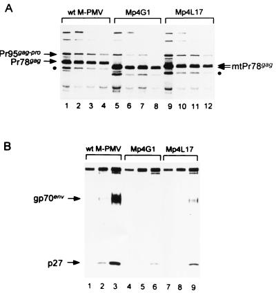
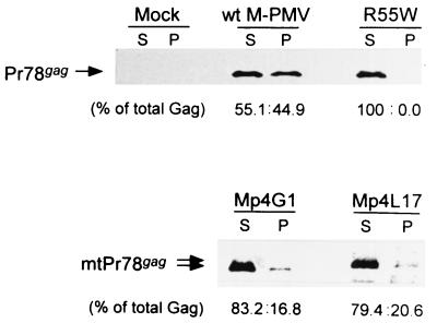
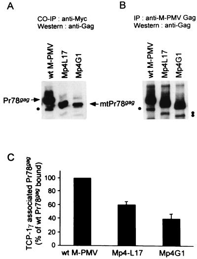
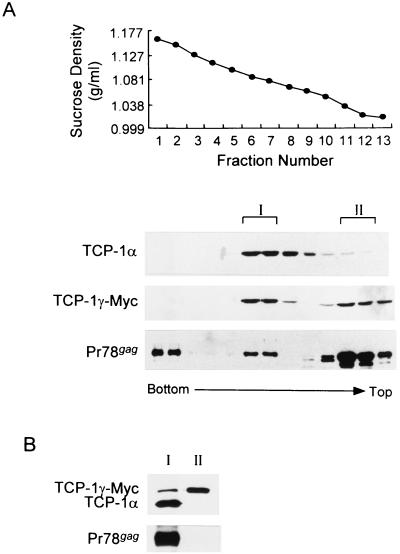
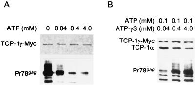
The carboxy terminus-encoding portion of the gag gene of Mason-Pfizer monkey virus (M-PMV), the prototype immunosuppressive primate type D retrovirus, encodes a 36-amino-acid, proline-rich protein domain that, in the mature virion, becomes the p4 capsid protein. The p4 domain has no known role in M-PMV replication. We found that two mutants with premature termination codons that remove half or all of the p4 domain produced lower levels of stable Gag protein and of self-assembled capsids. Interestingly, yeast two-hybrid screening revealed that p4 specifically interacted with TCP-1gamma, a subunit of the chaperonin TRiC (TCP-1 ring complex). TRiC is a cytosolic chaperonin that is known to be involved in both folding and subunit assembly of a variety of cellular proteins. TCP-1gamma also associated with high specificity with the M-PMV pp24/16-p12 domain and human immunodeficiency virus p6. Moreover, in cells, Gag polyprotein associated with the TRiC chaperonin complex and this association depended on ATP hydrolysis. In the p4 truncation mutants, the Gag-TRiC association was significantly reduced. These results strongly suggest that cytosolic chaperonin TRiC is involved in Gag folding and/or capsid assembly. We propose that TRiC associates transiently with nascent M-PMV Gag molecules to assist in their folding. Consequently, properly folded Gag molecules carry out the intermolecular interactions involved in self-assembly of the immature capsid.


Figures

FIG. 1
FIG. 1
Schematic representation of M-PMV mutants. The arrangement of the structural proteins within the Gag polyprotein is schematically presented (top). The partial amino acid sequences of the M-PMV p4 proteins are shown below, using single-letter amino acid codes. Prolines and carboxy-terminal residues are displayed, and other residues are indicated by dashed lines. Numerals indicate residue numbers. In Mp4L17, the 17th codon (TTA) was changed to a stop codon (TAA). In Mp4G1, the first codon (GGG) was changed to a stop codon (TAG). Arrowhead, cleavage site between the p14 NC protein and the p4 protein.
FIG. 2
FIG. 2
Immunoprecipitation of intracellular and extracellular viral proteins. To examine the biosynthesis and turnover of Gag (Pr78gag) and Gag-related precursor polyproteins (Pr95gag-pro), wild-type and mutant M-PMV proviral DNAs were transfected into COS-1 cells. (A) At 48 h after transfection, cells were pulse-labeled for 20 min with [3H]leucine (lanes 1, 5, and 9) and chased for 0.5 (lanes 2, 6, and 10), 1 (lanes 3, 7, and 11), and 2 h (lanes 4, 8, and 12). Cell-associated Gag-specific viral polyproteins were immunoprecipitated with rabbit anti-p27 CA antibody. An internal initiation product of the gag gene, Pr68gag, is marked (●). (B) Extracellular virions were pelleted from the culture medium of pulse-labeled cells after 0.5- (lanes 1, 4, and 7), 1- (lanes 2, 5, and 8), and 2-h (lanes 3, 6, and 9) chases and then immunoprecipitated with a goat anti-M-PMV antibody.
FIG. 3
FIG. 3
ICAP formation. To determine whether the p4-truncated Gag polyproteins can be assembled into capsids, ICAPs were prepared from lysates of HOS cells expressing wild-type M-PMV, the Mp4L17 and Mp4G1 truncation mutants, and a negative-control R55W mutant that is defective in ICAP assembly (36). Soluble (S) and capsid-associated pelletable (P) Gag polyproteins were fractionated and then immunoprecipitated with rabbit anti-Gag antiserum and visualized by Western blot assay with rabbit anti-Gag antibodies. The amount of Gag polyprotein in each band was quantified, and the ratios to the total amount of Gag protein (% of total Gag) were determined.
FIG. 4
FIG. 4
Coimmunoprecipitation of wild-type and p4-truncated Gag polyproteins with the TCP-1γ subunit. (A) The cellular interactions between M-PMV Gag polyprotein and TCP-1γ protein were examined by a coimmunoprecipitation (CO-IP) experiment. 293T cells transiently cotransfected with wild-type or p4 mutant (Mp4L17 or Mp4G1) proviral DNA and Myc-tagged TCP-1γ-expressing plasmid DNA were lysed in 1% NP-40-containing TNE buffer. TCP-1γ–Myc and its associated proteins were coimmunoprecipitated with mouse monoclonal anti-Myc antibody for 2 h at 4°C and then separated by 8%–SDS PAGE. TCP-1γ-Myc-bound Gag polyproteins were detected by Western blot assay with rabbit anti-Gag antibody. Dots, internal initiation products (Pr68gag). (B) To measure the total amount of Gag polyprotein in each sample, equal numbers of cotransfected 293T cells were lysed in lysis buffer A supplemented with 0.1% SDS and total Gag polyproteins were immunoprecipitated (IP) with rabbit anti-Gag antibody. Gag polyproteins were detected by Western blot assay with the same antibody. (C) The amount of Gag polyprotein in each band of panel A was quantified. The amount of TCP-1γ–Myc-bound Gag protein was normalized by that of total Gag (B) and then plotted as a percentage. The amount bound by wild-type (wt) M-PMV is arbitrarily presented as 100%. The data are the means and standard errors from three separate experiments.
FIG. 5
FIG. 5
Coimmunoprecipitation of M-PMV Gag polyprotein with the chaperonin TRiC. Cell fractionation and coimmunoprecipitation were carried out with 293T cells transiently cotransfected with M-PMV proviral DNA pSHRM15 and a Myc-tagged TCP-1γ-expressing plasmid. (A) The cells were lysed in 0.5% NP buffer and then fractionated through a continuous 5-to-40% (wt/vol) linear sucrose gradient. The fractions were collected from bottom to top and measured for sucrose density (top) Aliquots of each fraction were analyzed by Western blot assay with rat monoclonal anti-TCP-1α (TCP-1α), mouse monoclonal anti-Myc (TCP-1γ–Myc), and rabbit anti-Gag (Pr78gag). The densities of fractions 6 and 7 (1.076 and 1.086 g/ml, respectively) are near the expected density of the TRiC complex. (B) Peaks I and II (pooled fractions 6 and 7 and 11 and 12, respectively) were diluted with 0.5% NP buffer and then used to coimmunoprecipitate TCP-1γ–Myc and its associated proteins with a mouse monoclonal anti-Myc antibody. Proteins in the immune complex were analyzed as described above for TCP-1γ–Myc, TCP-1α, and Gag by Western blot assay.
FIG. 6
FIG. 6
Effects of ATP and ATP-γ-S on the M-PMV Gag association with the chaperonin TRiC. 293T cells transiently cotransfected with an M-PMV proviral DNA and a TCP-1γ–Myc-expressing plasmid were lysed in 1% NP-40-containing TNE buffer. (A) Cell lysates were incubated with 2 mM MgCl2 and various concentrations of ATP on ice for 1 h prior to coimmunoprecipitation with mouse monoclonal anti-Myc antibody. TCP-1γ–Myc and Gag proteins were detected by Western blot assay with mouse anti-Myc and rabbit anti-Gag antibodies, respectively. The sample with no ATP was sham treated with H2O. (B) To confirm the effect of ATP on dissociation of Gag proteins from TRiC, competition assays were carried out with nonhydrolyzable ATP analog ATP-γ-S. Cell lysates were treated for 1 h on ice with various concentrations of ATP-γ-S in the presence of 0.1 mM ATP and 2 mM MgCl2. After coimmunoprecipitation with an anti-Myc antibody, TCP-1γ–Myc, TCP-1α, and Gag proteins were detected as described above with mouse anti-Myc, rat anti-TCP-1α, and rabbit anti-Gag antibodies, respectively.

References

    1. Bachand F, Yao X J, Hrimech M, Rougeau N, Cohen É. Incorporation of Vpr into human immunodeficiency virus type 1 requires a direct interaction with the p6 domain of the p55 Gag precursor. J Biol Chem. 1999;274:9083–9091. - PubMed
    1. Bennett R P, Rhee S, Craven R C, Hunter E, Wills J W. Amino acids encoded downstream of gag are not required by Rous sarcoma virus protease during Gag-mediated assembly. J Virol. 1991;65:272–280. - PMC - PubMed
    1. Bradac J, Hunter E. Polypeptides of Mason-Pfizer monkey virus. I. Synthesis and processing of the gag-gene products. Virology. 1984;138:260–275. - PubMed
    1. Chattopadhyay S K, Sengupta D N, Fredrickson T N, Morse H C, Hartley J W. Characteristics and contributions of defective, ecotropic, and mink cell focus-inducing viruses involved in a retrovirus-induced immunodeficiency syndrome of mice. J Virol. 1991;65:4232–4241. - PMC - PubMed
    1. Chen C, Okayama H. High-efficiency transformation of mammalian cells by plasmid DNA. Mol Cell Biol. 1987;7:2745–2752. - PMC - PubMed

Publication types

MeSH terms