

Differences in cytokine and chemokine responses during neurological disease induced by polytropic murine retroviruses Map to separate regions of the viral envelope gene
- PMID: 11222710
- PMCID: PMC115911
- DOI: 10.1128/JVI.75.6.2848-2856.2001
Differences in cytokine and chemokine responses during neurological disease induced by polytropic murine retroviruses Map to separate regions of the viral envelope gene
Abstract
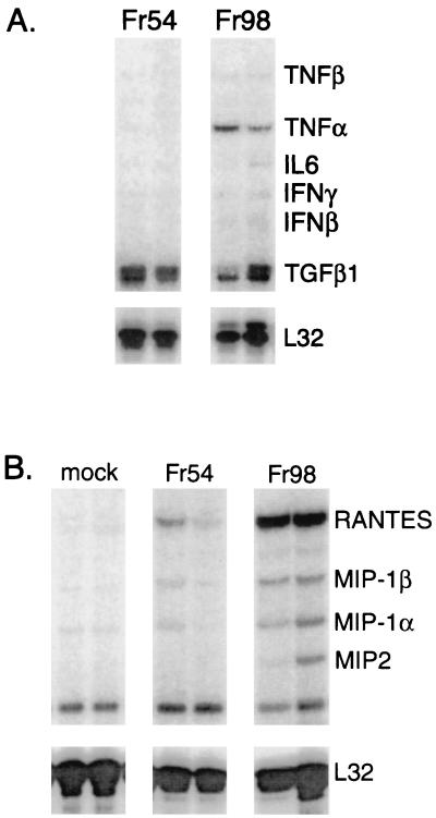
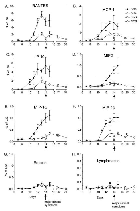
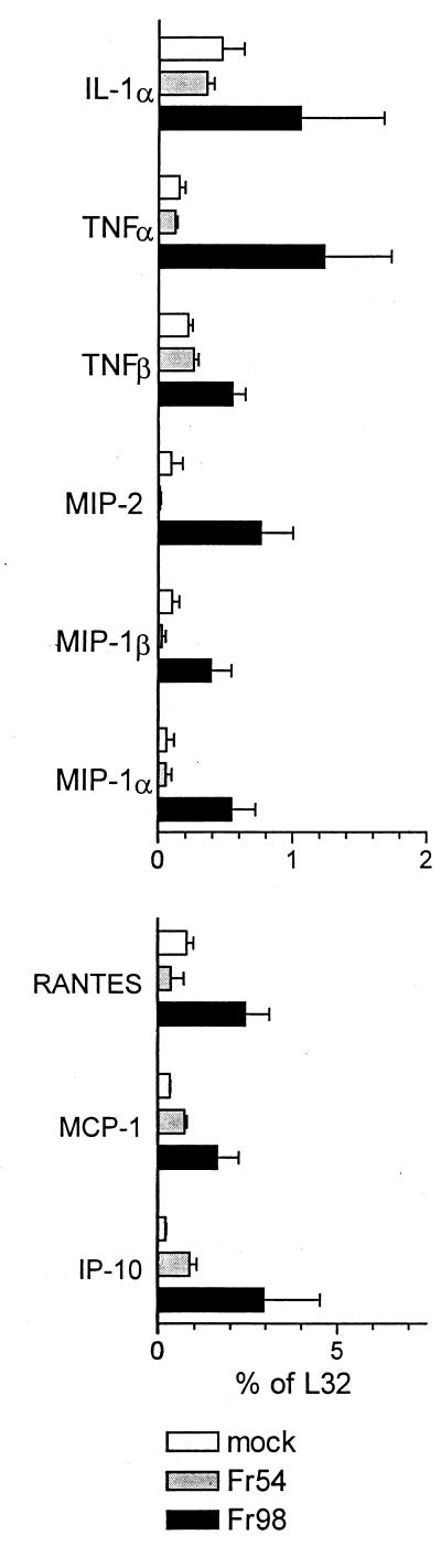
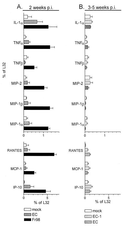
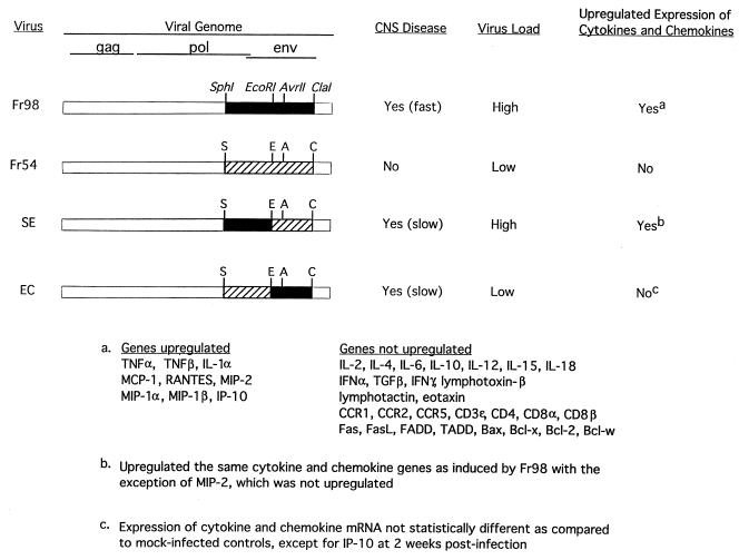
Infection of the central nervous system (CNS) by several viruses can lead to upregulation of proinflammatory cytokines and chemokines. In immunocompetent adults, these molecules induce prominent inflammatory infiltrates. However, with immunosuppressive retroviruses, such as human immunodeficiency virus (HIV), little CNS inflammation is observed yet proinflammatory cytokines and chemokines are still upregulated in some patients and may mediate pathogenesis. The present study examined expression of cytokines and chemokines in brain tissue of neonatal mice infected with virulent (Fr98) and avirulent (Fr54) polytropic murine retroviruses. While both viruses infect microglia and endothelia primarily in the white matter areas of the CNS, only Fr98 induces clinical CNS disease. The pathology consists of gliosis with minimal morphological changes and no inflammation, similar to HIV. In the present experiments, mice infected with Fr98 had increased cerebellar mRNA levels of proinflammatory cytokines tumor necrosis factor alpha (TNF-alpha), TNF-beta, and interleukin-1 alpha and chemokines macrophage inflammatory protein-1 alpha (MIP-1 alpha), MIP-1 beta, monocyte chemoattractant protein 1 (MCP-1), gamma-interferon-inducible protein 10 (IP-10), and RANTES compared to mice infected with Fr54 or mock-infected controls. The increased expression of these genes occurred prior to the development of clinical symptoms, suggesting that these cytokines and chemokines might be involved in induction of neuropathogenesis. Two separate regions of the Fr98 envelope gene are associated with neurovirulence. CNS disease associated with the N-terminal portion of the Fr98 env gene was preceded by upregulation of cytokines and chemokines. In contrast, disease associated with the central region of the Fr98 env gene showed no upregulation of cytokines or chemokines and thus did not require increased expression of these genes for disease induction.
Figures

Similar articles
-
Increased neurovirulence of polytropic mouse retroviruses delivered by inoculation of brain with infected neural stem cells.Virology. 1999 Oct 10;263(1):23-9. doi: 10.1006/viro.1999.9917. Virology. 1999. PMID: 10544079
-
Neurologic disease induced by polytropic murine retroviruses: neurovirulence determined by efficiency of spread to microglial cells.J Virol. 1997 Jul;71(7):5287-94. doi: 10.1128/JVI.71.7.5287-5294.1997. J Virol. 1997. PMID: 9188597 Free PMC article.
-
MCP-1 and CCR2 contribute to non-lymphocyte-mediated brain disease induced by Fr98 polytropic retrovirus infection in mice: role for astrocytes in retroviral neuropathogenesis.J Virol. 2004 Jun;78(12):6449-58. doi: 10.1128/JVI.78.12.6449-6458.2004. J Virol. 2004. PMID: 15163738 Free PMC article.
-
Influence of proinflammatory cytokines and chemokines on the neuropathogenesis of oncornavirus and immunosuppressive lentivirus infections.Curr Top Microbiol Immunol. 2006;303:67-95. doi: 10.1007/978-3-540-33397-5_4. Curr Top Microbiol Immunol. 2006. PMID: 16570857 Review.
-
Innate immunity in the pathogenesis of polytropic retrovirus infection in the central nervous system.Immunol Res. 2009;43(1-3):149-59. doi: 10.1007/s12026-008-8060-y. Immunol Res. 2009. PMID: 18818884 Free PMC article. Review.
Cited by
-
Neuroinvasion and Inflammation in Viral Central Nervous System Infections.Mediators Inflamm. 2016;2016:8562805. doi: 10.1155/2016/8562805. Epub 2016 May 25. Mediators Inflamm. 2016. PMID: 27313404 Free PMC article. Review.
-
Human endogenous retroviruses and multiple sclerosis: innocent bystanders or disease determinants?Biochim Biophys Acta. 2011 Feb;1812(2):162-76. doi: 10.1016/j.bbadis.2010.07.016. Epub 2010 Aug 6. Biochim Biophys Acta. 2011. PMID: 20696240 Free PMC article. Review.
-
Expression and function of chemokines during viral infections: from molecular mechanisms to in vivo function.J Leukoc Biol. 2003 Sep;74(3):331-43. doi: 10.1189/jlb.1102577. J Leukoc Biol. 2003. PMID: 12949236 Free PMC article. Review.
-
Endogenous retroviruses mobilized during friend murine leukemia virus infection.Virology. 2016 Dec;499:136-143. doi: 10.1016/j.virol.2016.07.009. Epub 2016 Sep 19. Virology. 2016. PMID: 27657834 Free PMC article.
-
Toll-like receptor 9-mediated protection of enterovirus 71 infection in mice is due to the release of danger-associated molecular patterns.J Virol. 2014 Oct;88(20):11658-70. doi: 10.1128/JVI.00867-14. Epub 2014 Jul 30. J Virol. 2014. PMID: 25078697 Free PMC article.
References
-
- Asensio V C, Campbell I L. Chemokines in the CNS: plurifunctional mediators in diverse states. Trends Neurosci. 1999;22:504–512. - PubMed
-
- Bacon K B, Harrison J K. Chemokines and their receptors in neurobiology: perspectives in physiology and homeostasis. J Neuroimmunol. 2000;104:92–97. - PubMed
-
- Basch R S, Grausz D, Harris N, Mitchison N A. Murine leukemia virus-associated cell surface antigens in rats neonatally infected with Gross murine leukemia virus. JNCI. 1979;63:1485–1492. - PubMed
-
- Boche D, Hurtrel M, Gray F, Claessens-Maire M A, Ganiere J P, Montagnier L, Hurtrel B. Virus load and neuropathology in the FIV model. J Neurovirol. 1996;2:377–387. - PubMed
MeSH terms
Substances
LinkOut - more resources
Full Text Sources
Other Literature Sources
Research Materials
Miscellaneous
