. 2001 Mar;75(6):2912-20.
doi: 10.1128/JVI.75.6.2912-2920.2001.

Inducible cyclic AMP early repressor produces reactivation of latent herpes simplex virus type 1 in neurons in vitro

Affiliations

Inducible cyclic AMP early repressor produces reactivation of latent herpes simplex virus type 1 in neurons in vitro

M A Colgin et al. J Virol. 2001 Mar.

Abstract

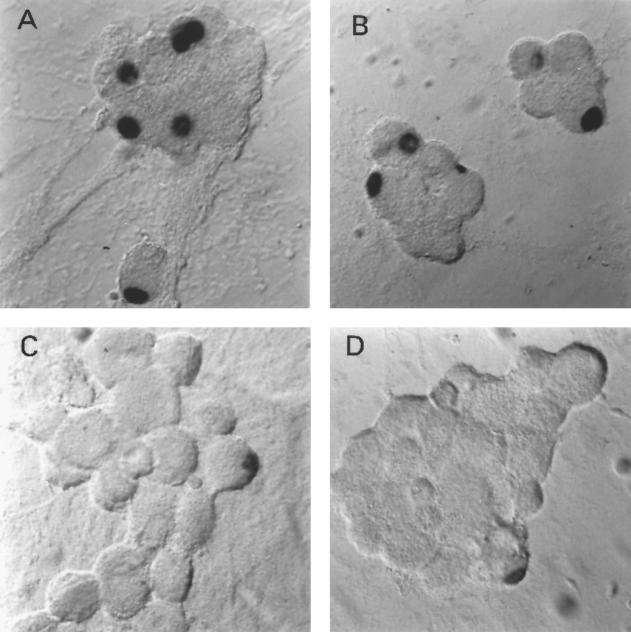
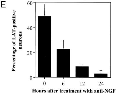
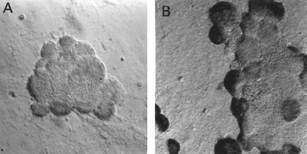
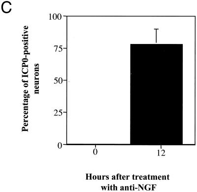
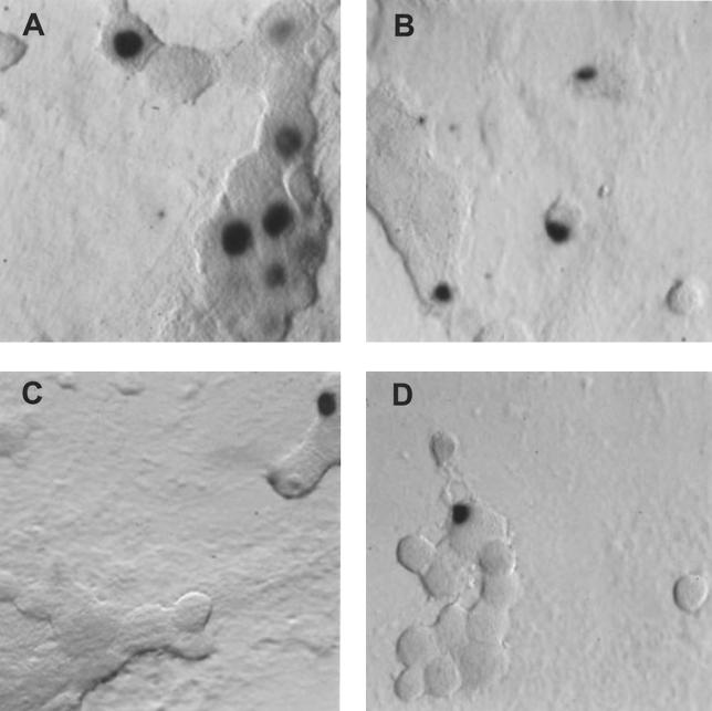
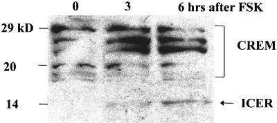
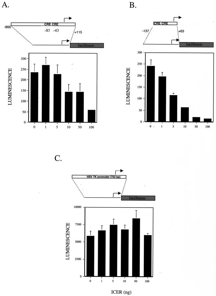
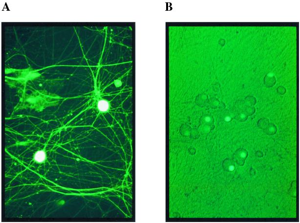
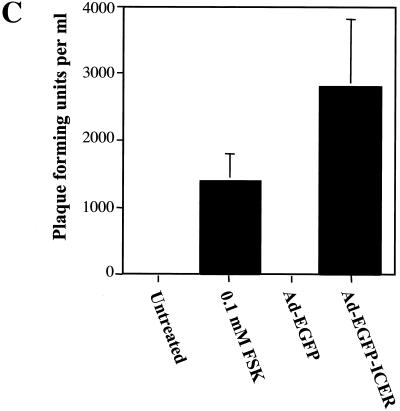
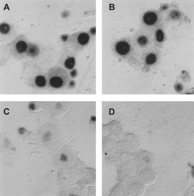
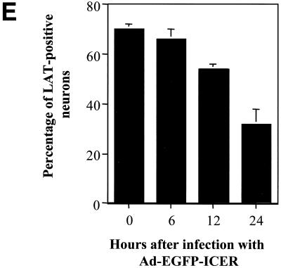
Herpes simplex virus type 1 (HSV-1) establishes a latent infection in neurons of the peripheral nervous system. During latent HSV-1 infection, viral gene expression is limited to latency-associated transcripts (LAT). HSV-1 remains latent until an unknown mechanism induces reactivation. The ability of the latent virus to periodically reactivate and be shed is essential to the transmission of disease. In vivo, the stimuli that induce reactivation of latent HSV-1 include stress, fever, and UV damage to the skin at the site of initial infection. In vitro, in primary neurons harboring latent HSV-1, nerve growth factor (NGF) deprivation or forskolin treatment induces reactivation. However, the mechanism involved in the induction of reactivation remains poorly understood. An in vitro neuronal model of HSV-1 latency was used to investigate potential mechanisms involved in the induction of reactivation of latent HSV-1. In situ hybridization analysis of neuronal cultures harboring latent HSV-1 showed a marked, rapid decrease in the percentage of LAT-positive neurons following induction of reactivation by NGF deprivation or forskolin treatment. Western blot analysis showed a corresponding increase in expression of the cellular transcription factor inducible cyclic AMP early repressor (ICER) during reactivation. In transient-transfection assays, ICER downregulated LAT promoter activity. Expression of ICER from a recombinant adenoviral vector induced reactivation and decreased the percentage of LAT-positive neurons in neuronal cultures harboring latent HSV-1. These results indicate that ICER represses LAT expression and induces reactivation of latent HSV-1.


Figures

FIG. 1
FIG. 1
In situ hybridization using a riboprobe to detect LAT in neuronal cultures latently infected with HSV-1 following the induction of reactivation by NGF deprivation. A digoxigenin-labeled riboprobe was used to detect LAT in neuronal cultures 2 weeks after the establishment of latency with HSV-1(F) following NGF deprivation. Representative fields are shown for cultures after induction of reactivation using NGF deprivation at 0 (A), 6 (B), 12 (C), and 24 (D) h after induction of reactivation. (E) Percentage of neurons per culture expressing LAT at the indicated time points. Values are means plus standard errors of the means (n = 4).
FIG. 1
FIG. 1
In situ hybridization using a riboprobe to detect LAT in neuronal cultures latently infected with HSV-1 following the induction of reactivation by NGF deprivation. A digoxigenin-labeled riboprobe was used to detect LAT in neuronal cultures 2 weeks after the establishment of latency with HSV-1(F) following NGF deprivation. Representative fields are shown for cultures after induction of reactivation using NGF deprivation at 0 (A), 6 (B), 12 (C), and 24 (D) h after induction of reactivation. (E) Percentage of neurons per culture expressing LAT at the indicated time points. Values are means plus standard errors of the means (n = 4).
FIG. 2
FIG. 2
In situ hybridization using a riboprobe to detect ICP0 in neuronal cultures latently infected with HSV-1 following the induction of reactivation by NGF deprivation. A digoxigenin-labeled riboprobe was used to detect ICP0 in neuronal cultures 2 weeks after the establishment of latency with HSV-1(F) following NGF deprivation. Representative fields are shown for cultures after induction of reactivation using NGF deprivation at 0 (A) and 12 (B) h after induction of reactivation. (C) Percentage of neurons per culture expressing ICP0 at the indicated time points. Values are means plus standard errors of the means (n = 4).
FIG. 2
FIG. 2
In situ hybridization using a riboprobe to detect ICP0 in neuronal cultures latently infected with HSV-1 following the induction of reactivation by NGF deprivation. A digoxigenin-labeled riboprobe was used to detect ICP0 in neuronal cultures 2 weeks after the establishment of latency with HSV-1(F) following NGF deprivation. Representative fields are shown for cultures after induction of reactivation using NGF deprivation at 0 (A) and 12 (B) h after induction of reactivation. (C) Percentage of neurons per culture expressing ICP0 at the indicated time points. Values are means plus standard errors of the means (n = 4).
FIG. 3
FIG. 3
In situ hybridization using a riboprobe to detect LAT in neuronal cultures latently infected with HSV-1 following treatment with forskolin (FSK). A digoxigenin-labeled riboprobe was used to detect LAT in neuronal cultures 2 weeks after establishment of latency with HSV-1(17+). Representative fields are shown for cultures after induction of reactivation using 0.5 mM forskolin at 0 (A), 6 (B), 12 (C), and 24 (D) h after induction of reactivation. (E) Percentage of neurons expressing LAT from the indicated time points. Values are means plus standard errors of the means (n = 4).
FIG. 3
FIG. 3
In situ hybridization using a riboprobe to detect LAT in neuronal cultures latently infected with HSV-1 following treatment with forskolin (FSK). A digoxigenin-labeled riboprobe was used to detect LAT in neuronal cultures 2 weeks after establishment of latency with HSV-1(17+). Representative fields are shown for cultures after induction of reactivation using 0.5 mM forskolin at 0 (A), 6 (B), 12 (C), and 24 (D) h after induction of reactivation. (E) Percentage of neurons expressing LAT from the indicated time points. Values are means plus standard errors of the means (n = 4).
FIG. 4
FIG. 4
Western blot analysis shows increased ICER in neuronal cultures latently infected with HSV-1 following treatment with forskolin (FSK). Neuronal cultures harboring latent HSV-1(17+) were treated with 0.1 mM forskolin. The anti-CREB primary antibody recognizes ICER as well as other CREM isoforms, producing a pattern of detected products similar to published results (21). The arrow indicates the predicted size of ICER.
FIG. 5
FIG. 5
Transient-transfection assays show that the ICER represses LAT promoter activity. Luciferase assays were performed to measure the ability of ICER to negatively regulate the LAT promoter activity. Vero cells were transiently transfected with 1 μg of reporter plasmid. Values are in luminescence units and are means plus standard deviations (n = 4). Cells were cotransfected with the indicated amount of ICER expression plasmid and the luciferase reporter containing plasmid. Luciferase expression from the full LAT promoter (A), the minimal LAT promoter (B), and the HSV thymidine kinase (TK) promoter (C) is shown.
FIG. 6
FIG. 6
Expression and induction of reactivation of latent HSV-1 in neurons using an adenoviral vector to express ICER fused to EGFP. Neurons in culture following infection with adenoviral vectors Ad-EGFP (A) and Ad-EGFP-ICER (B) showed expression of EGFP and EGFP-ICER, respectively. (C) Neuronal cultures, 2 weeks after establishment of latent HSV-1(17+) infections, were infected with the indicated adenoviral vector or treated with forskolin (FSK). Cultures were assayed for infectious virus 4 days posttreatment in plaque formation assays. Values are means plus standard errors of the means (n = 6).
FIG. 6
FIG. 6
Expression and induction of reactivation of latent HSV-1 in neurons using an adenoviral vector to express ICER fused to EGFP. Neurons in culture following infection with adenoviral vectors Ad-EGFP (A) and Ad-EGFP-ICER (B) showed expression of EGFP and EGFP-ICER, respectively. (C) Neuronal cultures, 2 weeks after establishment of latent HSV-1(17+) infections, were infected with the indicated adenoviral vector or treated with forskolin (FSK). Cultures were assayed for infectious virus 4 days posttreatment in plaque formation assays. Values are means plus standard errors of the means (n = 6).
FIG. 7
FIG. 7
In situ hybridization using a riboprobe to detect LAT in neuronal cultures latently infected with HSV-1 following infection with Ad-EGFP or Ad-EGFP-ICER. A digoxigenin-labeled riboprobe was used to detect LAT in neuronal cultures 2 weeks after the establishment of latent HSV-1(17+) following infection with adenoviral vectors. Fields shown are representative of neuronal cultures after infection with an adenoviral vector expressing EGFP at 24 h after infection (A) or an adenoviral vector expressing ICER-EGFP at 6 (B), 12 (C), and 24 (D) h after infection. (E) Percentage of neurons expressing LAT at the indicated time points. Values are means plus standard errors of the means (n = 4).
FIG. 7
FIG. 7
In situ hybridization using a riboprobe to detect LAT in neuronal cultures latently infected with HSV-1 following infection with Ad-EGFP or Ad-EGFP-ICER. A digoxigenin-labeled riboprobe was used to detect LAT in neuronal cultures 2 weeks after the establishment of latent HSV-1(17+) following infection with adenoviral vectors. Fields shown are representative of neuronal cultures after infection with an adenoviral vector expressing EGFP at 24 h after infection (A) or an adenoviral vector expressing ICER-EGFP at 6 (B), 12 (C), and 24 (D) h after infection. (E) Percentage of neurons expressing LAT at the indicated time points. Values are means plus standard errors of the means (n = 4).

Similar articles

Cited by

References

    1. Barnabas S, Hai T, Andrisani O M. The hepatitis B virus X protein enhances the DNA binding potential and transcription efficacy of bZip transcription factors. J Biol Chem. 1997;272:20684–20690. - PubMed
    1. Batchelor A H, O'Hare P. Regulation and cell-type-specific activity of a promoter located upstream of the latency-associated transcript of herpes simplex virus type 1. J Virol. 1990;64:3269–3279. - PMC - PubMed
    1. Block T M, Deshmane S, Masonis J, Maggioncalda J, Valyi-Nagi T, Fraser N W. An HSV null mutant reactivates slowly from latent infection and makes small plaques on CV-1 monolayers. Virology. 1993;192:618–630. - PubMed
    1. Bloom D C, Stevens J G, Hill J M, Tran R K. Mutagenesis of a cAMP response element within the latency-associated transcript promoter of HSV-1 reduces adrenergic reactivation. Virology. 1997;236:202–207. - PubMed
    1. Bodor J, Spetz A L, Strominger J L, Habener J F. cAMP inducibility of transcriptional repressor ICER in developing and mature human T lymphocytes. Proc Natl Acad Sci USA. 1996;93:3536–3541. - PMC - PubMed

Publication types