. 2001 Mar 1;29(5):1156-62.
doi: 10.1093/nar/29.5.1156.

Polysomal ribonuclease 1 exists in a latent form on polysomes prior to estrogen activation of mRNA decay

Affiliations

Polysomal ribonuclease 1 exists in a latent form on polysomes prior to estrogen activation of mRNA decay

K S Cunningham et al. Nucleic Acids Res. .

Abstract

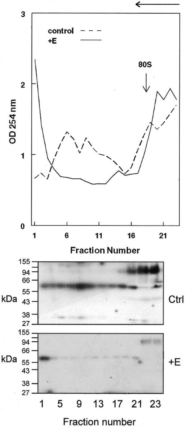
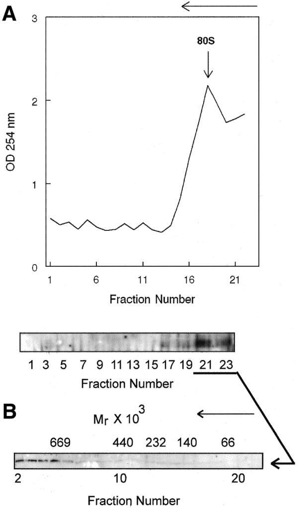
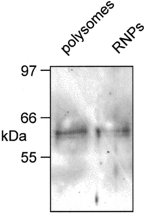
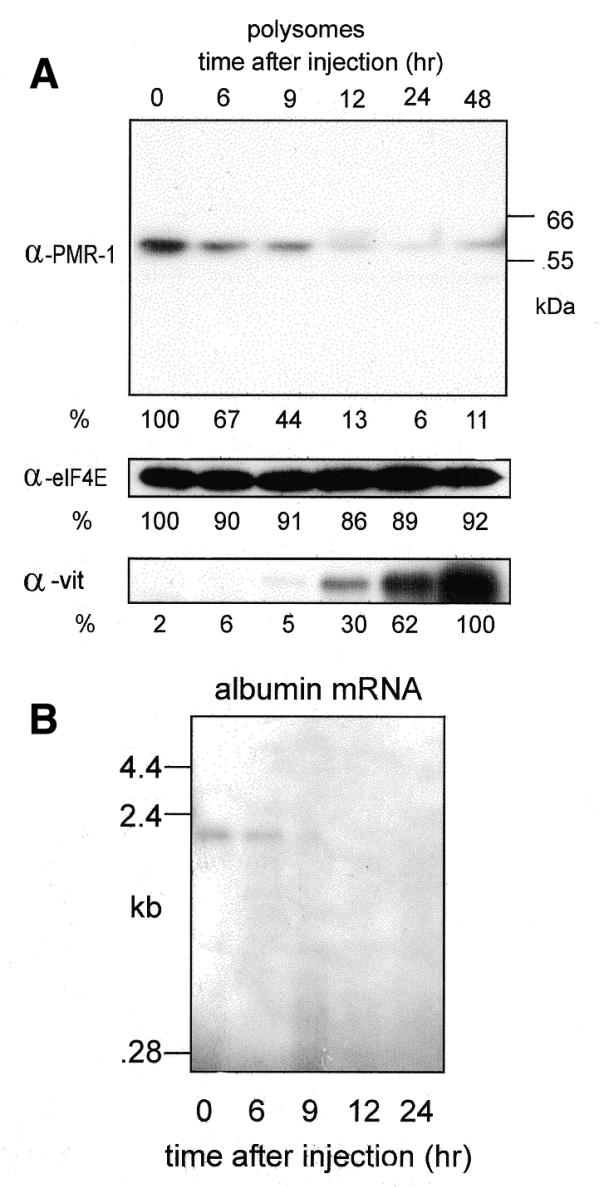
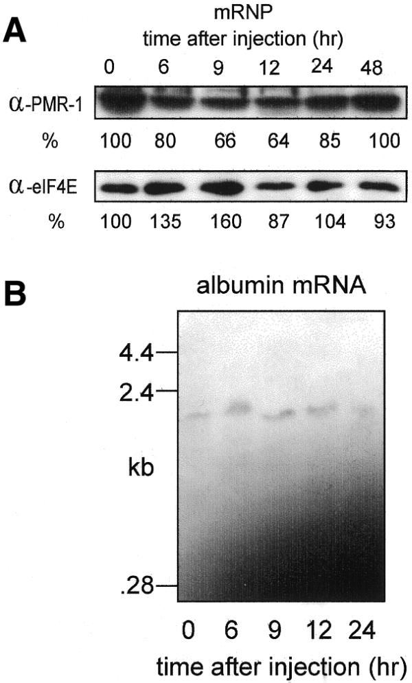
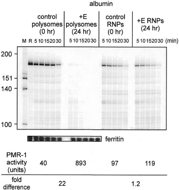
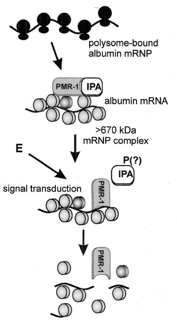
Estrogen induces a global change in the translation profile of Xenopus hepatocytes, replacing serum protein synthesis with production of the yolk protein precursor vitellogenin. This is accomplished by the coordinate destabilization of serum protein mRNAs and the transcriptional induction and subsequent stabilization of vitellogenin mRNA. Previous work identified an endonuclease activity whose appearance on polysomes correlated with the disappearance of serum protein mRNAs. This enzyme, polysomal ribonuclease 1 (PMR1), is a novel member of the peroxidase gene family. The current study examined the association of PMR1 with its mRNA targets on polysomes and mRNPs. The highest amount of polysome-bound PMR1 was observed prior to estrogen induction of mRNA decay. Its distribution on sucrose density gradients matched the absorbance profile of polysome-bound mRNA, suggesting that PMR1 forms a latent complex with mRNA. Following dissociation with EDTA the 62 kDa PMR1 sedimented with a larger complex of >670 kDa. Estrogen induces a 22-fold increase in unit enzymatic activity of polysome-bound PMR1, and a time-dependent loss of PMR1 from polysomes in a manner that mirrors the disappearance of albumin mRNA. These data suggest that the key step in the extensive estrogen-induced change in mRNA decay in Xenopus liver is activation of a latent mRNA endonuclease associated with its target mRNA.


Figures

Figure 1
Figure 1
Distribution of PMR1 on membrane-bound polysomes. Liver membrane-bound polysomes from control or 24 h estrogen-treated frogs were separated on a 20–50% sucrose density gradient and 0.5 ml fractions were collected at 4°C. The absorbance at 254 nm was determined for each fraction and is plotted in the top panel. The direction of sedimentation is indicated by the arrow at the top of the diagram. In the bottom panel 30 µl of each odd numbered sample was analyzed by western blot using a polyclonal antibody to PMR1. The position of 80S monoribosome complexes is shown by an arrow.
Figure 2
Figure 2
Glycerol gradient analysis of polysome-bound PMR1. (A) Membrane-bound polysomes prepared from control frogs were incubated for 45 min with gentle rocking at 4°C with 50 mM EDTA to dissociate ribosome-bound complexes, followed by fractionation on a 20–50% sucrose density gradient. The absorbance at 254 nm was determined for each fraction and is plotted in the top panel. In the bottom panel odd numbered samples were analyzed by western blot as in Figure 1 to identify PMR1-containing fractions. (B) Fractions 21–23 from the sucrose gradient in (A) were pooled, dialyzed and applied to a 10–40% glycerol gradient. Fractions obtained after centrifugation for 20 h at 83 000 gmax were analyzed by western blot as in (A). Molecular size markers consisting of a mixture of bovine serum albumin (Mr = 66 000), lactate dehydrogenase (Mr = 140 000), catalase (Mr = 232 000), ferritin (Mr = 440 000) and thyroglobulin (Mr = 669 000) were fractionated on a parallel gradient. Their sedimentation positions are indicated above the autoradiogram. The direction of sedimentation is indicated by an arrow above each figure.
Figure 3
Figure 3
Recovery of PMR1 on oligo(dT)–cellulose. Post-nuclear liver extract prepared from 5 g liver from control frogs was fractionated on a discontinuous sucrose step gradient to prepare mRNPs and polysomes (20). These were bound to oligo(dT)–cellulose for 2 h at 4°C and the matrix was washed and eluted with water. The recovered material was dissolved in 20 µl of 10 mM Tris–HCl, pH 8.0, RNA was removed by digestion for 10 min at 37°C with 5 µg of RNase A and the sample was assayed by western blot for PMR1.
Figure 4
Figure 4
Estrogen-induced changes in polysome-bound PMR1. (A) Polysomes were prepared at the times indicated in the figure and normalized to 100 µg protein. These were separated by SDS–PAGE and analyzed by western blot using antibodies to PMR1, eIF4E and vitellogenin. The positions of the 55 and 66 kDa size markers are indicated in the PMR1 western. The relative signal intensity of each band was determined by scanning densitometry. For all but vitellogenin the signal at time 0 was arbitrarily set to 100%. For vitellogenin the maximal signal at 48 h was arbitrarily set to 100%. (B) Five micrograms of total RNA from each of the polysome fractions examined in (A) was analyzed by northern blot using an antisense probe for albumin mRNA.
Figure 5
Figure 5
Estrogen-induced changes in mRNP-bound PMR1. (A) Samples of the mRNP fraction prepared from the same animals as in Figure 4 were normalized to 100 µg protein, separated by SDS–PAGE and analyzed by western blot as in Figure 4 using antibodies to PMR1 and eIF4E. The stronger PMR1 signal intensity seen here is a result of a longer exposure time and does not reflect the relative amount of PMR1 in these samples. mRNPs at time 0 contain 10% as much PMR1 as polysomes. (B) Five micrograms of total RNA from each of the polysome fractions examined in (A) was analyzed by northern blot using an antisense probe for albumin mRNA.
Figure 6
Figure 6
Estrogen-induced increase in PMR1 unit activity. Polysomes and mRNP complexes isolated from control and 24 h estrogen-treated frogs were normalized by scanning densitometry to equal amounts of immunoreactive PMR1. These were incubated with a uniformly 32P-labeled 160 nt albumin transcript (18) or a full-length 32P-labeled ferritin transcript for the indicated times and separated on a denaturing polyacrylamide–urea gel. The input transcript is denoted R in lane 2. The units of PMR1 enzymatic activity were determined by phosphorimager analysis of data obtained from the resulting albumin RNA decay curves. One unit of PMR1 activity equals the amount of enzyme that completely degrades 7 fmol albumin transcript substrate in 30 min at 22°C (17).
Figure 7
Figure 7
A model for activation of mRNA decay by PMR1. In this model PMR1 present on polysomes as part of a large RNP complex is maintained in a latent form by a bound inhibitory protein IPA (inhibitor of PMRI activity). Estrogen (E) activates a signal transduction pathway resulting in post-translational modification of IPA (indicated here as phosphorylation, but that is only speculation), causing it to dissociate from PMR1 and activate cleavage of albumin mRNA within the complex.

References

    1. Ross J. (1996) Control of messenger RNA stability in higher eukaryotes. Trends. Genet., 12, 171–175. - PubMed
    1. Jacobson A. and Peltz,S.W. (1996) Interrelationships of the pathways of mRNA decay and translation in eukaryotic cells. Annu. Rev. Biochem., 65, 693–739. - PubMed
    1. Tharun S. and Parker,R. (1997) Mechanisms of mRNA turnover in eukaryotic cells. In Harford,J. and Morris,D.R. (eds), mRNA Metabolism and Post-transcriptional Gene Regulation. Wiley, New York, NY, pp. 181–200.
    1. Couttet P., Fromont-Racine,M., Steel,D.M., Pictet,R. and Grange,T. (1997) Messenger RNA deadenylation precedes decapping in mammalian cells. Proc. Natl Acad. Sci. USA, 94, 5628–5633. - PMC - PubMed
    1. Tarun S.Z.,Jr, Wells,S.E., Deardorff,J.A. and Sachs,A.B. (1997) Translation initiation factor eIF4G mediates in vitro poly(A) tail-dependent translation. Proc. Natl Acad. Sci. USA, 94, 9046–9051. - PMC - PubMed

Publication types