. 2001 Jan 15;20(1-2):40-6.
doi: 10.1093/emboj/20.1.40.

Structure of a genetically engineered molecular motor

Affiliations

Structure of a genetically engineered molecular motor

W Kliche et al. EMBO J. .

Abstract

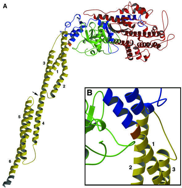
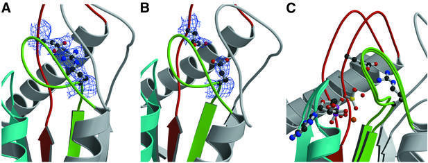
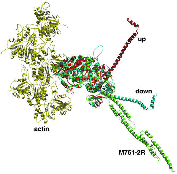
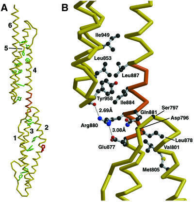
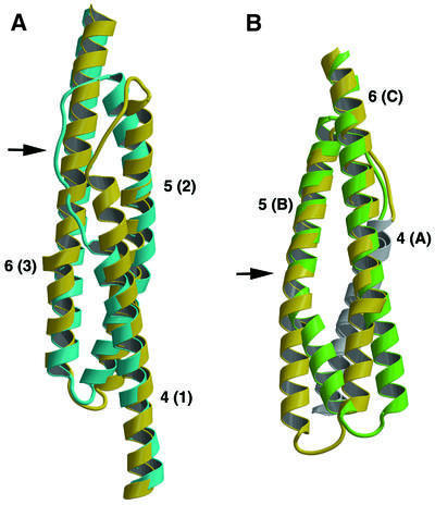
Molecular motors move unidirectionally along polymer tracks, producing movement and force in an ATP-dependent fashion. They achieve this by amplifying small conformational changes in the nucleotide-binding region into force-generating movements of larger protein domains. We present the 2.8 A resolution crystal structure of an artificial actin-based motor. By combining the catalytic domain of myosin II with a 130 A conformational amplifier consisting of repeats 1 and 2 of alpha-actinin, we demonstrate that it is possible to genetically engineer single-polypeptide molecular motors with precisely defined lever arm lengths and specific motile properties. Furthermore, our structure shows the consequences of mutating a conserved salt bridge in the nucleotide-binding region. Disruption of this salt bridge, which is known to severely inhibit ATP hydrolysis activity, appears to interfere with formation of myosin's catalytically active 'closed' conformation. Finally, we describe the structure of alpha-actinin repeats 1 and 2 as being composed of two rigid, triple-helical bundles linked by an uninterrupted alpha-helix. This fold is very similar to the previously described structures of alpha-actinin repeats 2 and 3, and alpha-spectrin repeats 16 and 17.


Figures

None
Fig. 1. Structure of M761-2R-R238E. Although two molecules are present in the crystallographic asymmetric unit, only one is shown here. The two molecules are essentially identical throughout the myosin motor domain (residues 2–761). However, upon leaving the converter domain, the lever arms assume slightly different orientations and deviate at the ends by 19.4 Å. (A) A complete molecule spanning amino acids 2–1010 is shown. No electron density was observed for five residues at the N-terminus, the loop region 205–208 and one residue at the C-terminus. The N-terminal domain (2–200) is shown in green; 50 kDa domain in red (201–613); C-terminal and converter domain in blue (614–761); linker region in orange (762–764); α-actinin lever arm in yellow (765–1003); and seven histidines from the His8 purification tag in gray (1004–1010). The linker region is composed of three residues (Leu-Gly-Arg) introduced during cloning. The observed lever arm is ∼140 Å long (measured from Cα of 761 to Cα of 1010). Each α-actinin repeat contributes ∼65 Å, and the histidine purification tag another 10 Å. Helices 1–3 make up the first α-actinin repeat, and 4–6 the second. The arrowhead indicates the α-helical region linking the two repeats. The disruptive kink in helix 2 is caused by the presence of two adjacent proline residues (see Figure 4A). (B) Detailed view of the linker region joining the myosin converter domain to helix 1 of α-actinin. The view is rotated 180° around a vertical axis from that in (A).
None
Fig. 2. Detailed view of the conserved salt bridge linking switch I and switch II. The conserved nucleotide binding/sensing elements found in all myosins, kinesins and G-proteins are highlighted for the P-loop in blue, switch I in green and switch II in red. (A) The structure of Dictyostelium myosin II motor complexed with Mg-ADP-BeF3 (F.J.Kull and K.C.Holmes, unpublished results). As in Mg-ADP-VO4 (Smith and Rayment, 1996) and Mg-ADP-BeF3 (Dominguez et al., 1998) structures, switch I and switch II are closed. The conserved salt bridge between residues R238 and E459 is shown as a ball-and-stick model surrounded by 2.6 Å experimental 2FoFc electron density (blue wire-frame), contoured at 1σ. As expected for a salt bridge, the electron density is continuous between the residues, which point toward each other. (B) The same region as observed in the crystal structure of M761-2R-R238E. The electron density was calculated from a model with alanines at positions 238 and 459 in order to eliminate model bias. Electron density for two glutamic acid residues is clearly visible, but the side chain of E238 now points away from E459 and the switch II loop has moved away from switch I. (C) The same region showing a superposition of the M761-2R-R238E structure with a structure of Dictyostelium myosin II motor complexed with Mg-ADP-VO4 (PDB code 1VOM) (Smith and Rayment, 1996). The nucleotide and R238–E459 salt bridge are shown as ball-and-stick models. Both the P-loop and switch I regions are in essentially identical conformations in both structures. However, the switch II region (red) shifts to the right, towards the nucleotide, by ∼5 Å in the Mg-ADP-VO4 structure, allowing the formation of the R238–E459 salt bridge.
None
Fig. 3. Orientation of the myosin lever arm. Five molecules of actin making up part of a helical actin filament are shown in yellow. Modeled on to this are myosin in the ‘pre-power-stroke’ up/closed orientation in red, the ‘post-power-stroke’ down/open orientation in blue, and the M761-2R-R238E structure in green. The up, down and actomyosin complex structures were modeled as described previously (Geeves and Holmes, 1999). The M761-2R-R238E structure was then aligned to the core domain of the down/open structure via residues 160–200, which includes the highly conserved P-loop region. Note that in the M761-2R-R238E structure, the helix leaving the converter domain initially superposes with the down/open structure, but then deviates due to the different helical bend of the α-actinin.
None
Fig. 4. The structure of α-actinin repeats 1 and 2. (A) An α-carbon chain trace of the six helices making up repeats 1 (helices labeled 1–3) and 2 (helices labeled 4–6) is shown in yellow. The 17 hydrophobic aromatic amino acid residues stabilizing the triple-helical packing are shown in green (seven tyrosines, six phenylalanines and four tryptophans). Two adjacent proline residues are shown in red, which cause a kink but not a break in α-helix 2 of repeat 1. The uninterrupted α-helix linking repeats 1 and 2 is shown in orange. (B) Detailed view of the linker region, highlighting the stabilizing hydrophobic and hydrogen bonding interactions. Colors and orientation are identical to those in (A). Side chains are shown as ball-and-stick models, with the exception of Asp796 and Ser797, in which only the α-carbon atoms involved in hydrophobic contacts are shown for clarity. The salt bridge between Arg880 and Glu877, and the hydrogen bond between Arg880 and the carbonyl oxygen of Leu956 (also shown as a ball-and-stick model), are shown as dashed lines.
None
Fig. 5. Comparison of Dictyostelium α-actinin with human α-actinin and human α-spectrin. (A) The overlapping repeat 2 region of Dictyostelium (yellow) and human (blue) α-actinin are shown as ribbon diagrams. Helices are numbered as described above for Dictyostelium α-actinin and, in parentheses, as described previously for human α-actinin (Djinovic-Carugo et al., 1999). The largest differences occur in the loop region connecting helices 4 and 5, indicated by an arrow, where the human α-actinin structure would seriously overlap with Dictyostelium helix 6. (B) The alignment of Dictyostelium repeat 2 (yellow) with repeat 16 human α-spectrin (green) is shown as ribbon diagrams. Helices are numbered as described above for the Dictyostelium protein and, in parentheses, as described previously for the human protein (Grum et al., 1999). Dictyostelium helix 4 and α-spectrin helix A, which are in the background, are colored white for clarity. In general, the two structures align more closely than the human/Dictyostelium alignment described above. The largest difference occurs in the loop region connecting helices 5 and 6, indicated by an arrow, where the human α-spectrin structure is moved in respect to the Dictyostelium α-actinin structure as a result of a proline-induced kink in helix B.

References

    1. Anson M., Geeves,M.A., Kurzawa,S.E. and Manstein,D.J. (1996) Myosin motors with artificial lever arms. EMBO J., 15, 6069–6074. - PMC - PubMed
    1. Blanchard A., Ohanian,V. and Critchley,D. (1989) The structure and function of α-actinin. J. Muscle Res. Cell Motil., 10, 280–289. - PubMed
    1. Brunger A.T. et al. (1998) Crystallography and NMR system (CNS): a new software system for macromolecular structure determination. Acta Crystallogr. D, 54, 905–921. - PubMed
    1. Burley S.K. et al (1999) Structural genomics: beyond the human genome project. Nature Genet., 23, 151–157. - PubMed
    1. Corrie J.E. et al. (1999) Dynamic measurement of myosin light-chain-domain tilt and twist in muscle contraction. Nature, 400, 425–430. - PubMed

Publication types