. 2001 Jan 15;20(1-2):101-8.
doi: 10.1093/emboj/20.1.101.

Coiled-coil domain-mediated FRQ-FRQ interaction is essential for its circadian clock function in Neurospora

Affiliations

Coiled-coil domain-mediated FRQ-FRQ interaction is essential for its circadian clock function in Neurospora

P Cheng et al. EMBO J. .

Abstract

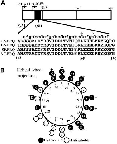
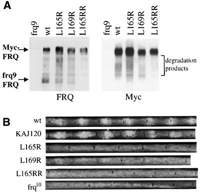
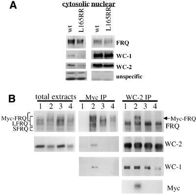
The frequency (frq) gene, the central component of the frq-based circadian negative feedback loop, regulates various aspects of the circadian clock in NEUROSPORA: However, the biochemical function of its protein products, FRQ, is poorly understood. In this study, we demonstrated that the most conserved region of FRQ forms a coiled-coil domain. FRQ interacts with itself in vivo, and the deletion of the coiled-coil region results in loss of the interaction. Point mutations, which are designed to disrupt the coiled-coil structure, weaken or completely abolish the FRQ self-association and lead to the arrhythmicity of the overt rhythm. Mutations of the FRQ coiled-coil that inhibit self-association also prevent its interaction with two other key components of the NEUROSPORA: circadian clock, namely WC-1 and WC-2, the two PAS domain-containing transcription factors. Taken together, these data strongly suggest that the formation of the FRQ-FRQ and FRQ-WC complexes is essential for the function of the NEUROSPORA: clock.


Figures

None
Fig. 1. The putative FRQ coiled-coil domain. (A) Schematic diagram of the FRQ ORF and sequencing alignments of the putative FRQ coiled-coil domain from four different fungal species. The open box represents the FRQ ORF. Arrows labeled AUG#1 and AUG#3 are the two alternative protein initiation sites that produce LFRQ and SFRQ. The filled black box represents the putative FRQ coiled-coil domain. NLS, nuclear localization signal. The line in the middle represents the location of the frq9 mutation. In the sequence alignments, the bold letters are the conserved amino acids in different frq homologs. On the top of the alignment, letters ag label the position of each amino acid within the helical heptad repeats. Asterisks mark the two amino acids (Leu165 and Leu169) that are mutated in this study. CS, Chromocrea spinlosa; LA, Leptosphaeria australiensis; SF, Sordaria fimicola; NC, Neurospora crassa. (B) Helical wheel projection of the FRQ coiled-coil domain. In the coiled-coils, seven residues allow the helix to make two turns to show a heptad repeat. The Ser145 denotes as position ‘1’ in the helical wheel projection. The charge and the hydrophobicity of each residue are labeled.
None
Fig. 2. The expression of Myc-FRQ in Neurospora and the rescue of the conidiation banding rhythm of the frq null strain by Myc-FRQ. (A) Western blot analysis showing the expression profile of Myc-FRQ in Neurospora. pKAJ120⋅Myc-FRQ was transformed into 93–4 (frq null strain) and seven independent transformants were selected for protein analysis. Cultures were grown and harvested in constant light (LL). The protein blot was first probed with FRQ antiserum (top) and then stripped and reprobed with a monoclonal c-Myc antibody (bottom). Note the slower mobility of Myc-FRQ bands relative to the wild-type FRQ. (B) The rescue of the circadian conidiation banding rhythm of frq null strain by Myc-FRQ. Both the original image of the race tubes and the densitometric analysis of the race tubes are shown. frq10 is the frq null strain. The race tubes shown are representative samples from six replicate tubes. (C) Expression of Myc-FRQ in an frq+ strain. pKAJ120⋅Myc-FRQ was transformed into 87–12 (frq+) and two independent transformants (1 and 2) are analyzed along with the wild-type strain. The blot was probed with either FRQ (upper panel) or c-Myc antibody (lower panel).
None
Fig. 3. FRQ interacts with itself in vivo. (A) Western blot analyses (probed with FRQ or c-Myc antibody) showing the expression of various FRQ forms in total extracts (left two panels) and in immunoprecipitates (right two panels). Immunoprecipitation was performed using a c-Myc monoclonal antibody. Note the lack of low molecular weight smear in the right bottom blot probed with c-Myc antibody. Lane 1, frq+, Myc-FRQ; LL; lane 2, wild type, LL; lane 3, frq+, Myc-FRQ; DD20; lane 4, wild type, DD20. (B) Phosphatase treatment after immunoprecipitation shows that Myc-FRQ interacts with the endogenous FRQ forms. Before phosphatase treatment, the protein extracts were immunoprecipitated with either FRQ (center panel) or c-Myc (right panel) antibodies. The cultures were grown and harvested in LL. Lane 1, wild type; lane 2, frq+, Myc-FRQ.
None
Fig. 4. Deletion of the putative coiled-coil domain abolishes FRQ–FRQ interaction. (A) The interaction between Myc-FRQ and the truncated frq9 FRQ. The cultures were grown in LL. Lane 1, frq10 (frq null); lane 2, frq9; lane 3, frq9, Myc-FRQ; lane 4, frq9, Myc-FRQ1 (deletion of the coiled-coil region). The left four lanes are total protein extracts and the three lanes on the right are IP products with c-Myc antibody. The protein blots were probed with FRQ or c-Myc antibody. Note the disappearance of the frq9 FRQ bands in lane 4 of the IP samples. (B) Myc-FRQ with the deletion of the coiled-coil domain can no longer interact with the endogenous FRQ forms. Lane 1, wild type; lane 2, frq+, Myc-FRQ; lane 3, frq+, Myc-FRQ1. The protein extracts were immunoprecipitated with FRQ (center) or c-Myc (right) antibody and then treated with phosphatase to dephosphorylate various FRQ forms. The protein blots were probed with FRQ antiserum. Note the disappearance of the LFRQ and SFRQ bands in lane 3 of the Myc IP panel.
None
Fig. 5. Point mutations of the coiled-coil domain lead to the weakening or complete loss of the FRQ–FRQ interaction and loss of rescue of the conidiation banding rhythm in the frq null strain. (A) Leu165 and Leu169 are important residues for FRQ–FRQ interaction. Single (Leu165Arg and Leu169Arg) or double (Leu165ArgArg) mutations were introduced into pKAJ120⋅Myc-FRQ. Mutation constructs were transformed into 94–1 (frq9, his-3), so that both Myc-FRQ and frq9 FRQ are expressed in these transformants. Various Myc-FRQ forms were immunoprecipitated down using c-Myc antibody. The protein blots were probed with FRQ or Myc antibody. Note the decrease of the frq9 FRQ signals in Leu165Arg and Leu169Arg samples, and its complete disappearance in 165ArgArg sample. The smearing signals between Myc-FRQ and frq9 FRQ are degradation products of Myc-FRQ. (B) Race tube results showing the arrhythmicity in constant darkness for the FRQ coiled-coil mutant strains. Leu165Arg, Leu169Arg and Leu165ArgArg mutations were introduced into pKAJ120. The mutations constructs were then transformed into the frq null strain (frq10). The resulting transformants were first screened by western blot analysis for FRQ expression before being examined by race tube assays. At least 10 positive transformants were examined by race tube assays. The wild-type strain and the pKAJ120 transformants were used as controls. The race tubes shown are representative samples from six replicate tubes.
None
Fig. 6. The FRQ coiled-coil domain is not required for the nuclear localization of FRQ but it is necessary for the FRQ–WC interactions. (A) Western blot analysis of the nuclear and cytosolic preparations shows that the coiled-coil mutation does not affect the nuclear localization of FRQ. Wild-type strain and Leu165ArgArg mutant strain (frq10, pKAJ120⋅L165RR) were grown and harvested in LL. The protein blots were probed with either FRQ, WC-1 or WC-2 antiserum. The two non-specific bands (bottom panels), recognized by the WC-2 antiserum, can only be found in the cytosolic fractions, suggesting that the nuclei preparations are free of cytosolic contamination. (B) The formation of the FRQ coiled-coil structure is required for the interaction between FRQ and the WC proteins. Total protein extracts from different strains grown in LL were immunoprecipitated by c-Myc or WC-2 antibody, and protein blots were probed with FRQ, WC-1, WC-2 or c-Myc antibody. Lane 1, frq+; lane 2, frq+, Myc-FRQ; lane 3, frq+, Myc-FRQ1; lane 4, frq+, FRQ-165RR.

References

    1. Allada R., White,N.E., So,W.V., Hall,J.C. and Rosbash,M. (1998) A mutant Drosophila homolog of mammalian Clock disrupts circadian rhythms and transcription of period and timeless. Cell, 93, 791–804. - PubMed
    1. Aronson B., Johnson,K., Loros,J.J. and Dunlap,J.C. (1994a) Negative feedback defining a circadian clock: autoregulation in the clock gene frequency. Science, 263, 1578–1584. - PubMed
    1. Aronson B.D., Johnson,K.A. and Dunlap,J.C. (1994b) The circadian clock locus frequency: A single ORF defines period length and temperature compensation. Proc. Natl Acad. Sci. USA, 91, 7683–7687. - PMC - PubMed
    1. Bell-Pedersen D., Shinohara,M., Loros,J. and Dunlap,J.C. (1996) Circadian clock-controlled genes isolated from Neurospora crassa are late night to early morning specific. Proc. Natl Acad. Sci. USA, 93, 13096–13101. - PMC - PubMed
    1. Berger B., Wilson,D.B., Wolf,E., Tonchev,T., Milla,M. and Kim,P.S. (1995) Predicting coiled coils by use of pairwise residue correlations. Proc. Natl Acad. Sci. USA, 92, 8259–8263. - PMC - PubMed

Publication types

MeSH terms