. 2001 Feb;13(2):303-18.
doi: 10.1105/tpc.13.2.303.

Wall-associated kinases are expressed throughout plant development and are required for cell expansion

Affiliations

Wall-associated kinases are expressed throughout plant development and are required for cell expansion

T A Wagner et al. Plant Cell. 2001 Feb.

Abstract

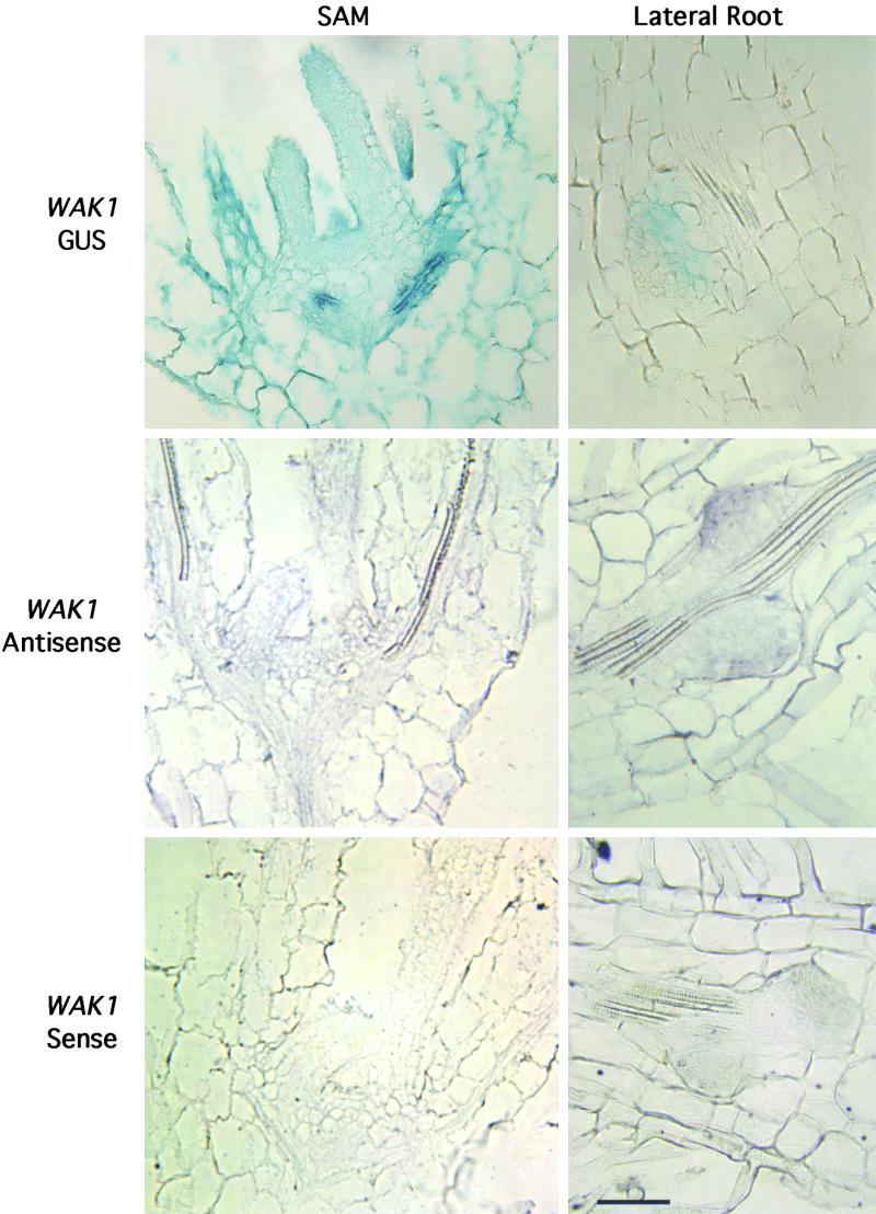
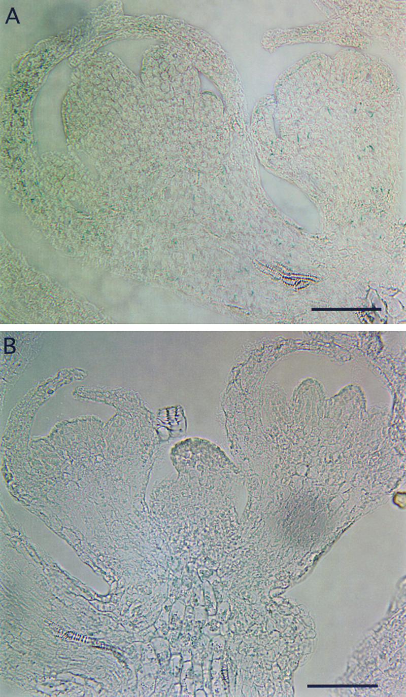
The mechanism by which events in the angiosperm cell wall are communicated to the cytoplasm is not well characterized. A family of five Arabidopsis wall-associated kinases (WAKs) have the potential to provide a physical and signaling continuum between the cell wall and the cytoplasm. The WAKs have an active cytoplasmic protein kinase domain, span the plasma membrane, and contain an N terminus that binds the cell wall. We show here that WAKs are expressed at organ junctions, in shoot and root apical meristems, in expanding leaves, and in response to wall disturbances. Leaves expressing an antisense WAK gene have reduced WAK protein levels and exhibit a loss of cell expansion. WAKs are covalently bound to pectin in the cell wall, providing evidence that the binding of a structural carbohydrate by a receptor-like kinase may have significance in the control of cell expansion.


Figures

Figure 1.
Figure 1.
WAK1and WAK2 Promoters Are Differentially Active in Seedlings. T3 seedlings containing the WAK1 promoter–GUS transgene (at left) or the WAK2 promoter–GUS transgene (at right ) were grown on plates for 1, 2, and 3 days in the growth chamber and then stained for GUS.
Figure 2.
Figure 2.
WAK1 Is Expressed in Root and Shoot Meristems. Sections through 7-day-old seedlings containing the WAK1 promoter–GUS transgene and stained for GUS (top) were compared with similar sections that were probed with the antisense (middle) and sense (bottom) WAK1 transcript. formula image.
Figure 3.
Figure 3.
WAK2 Is Expressed in Root and Shoot Meristems. Sections through 7-day-old seedlings containing the WAK2 promoter–GUS transgene and stained for GUS (top) were compared with similar sections that were probed with the antisense (middle) and sense (bottom) WAK2 transcript. formula image.
Figure 4.
Figure 4.
The WAK1, WAK2, and WAK3 Promoters Are Active in Adult-Stage Plants. (A) to (C) Young rosette plants grown in soil for 14 days. (A) WAK1 promoter–GUS; (B) WAK2 promoter–GUS; and (C) WAK3 promoter–GUS. (D) and (E) GUS staining at the node of the inflorescense stem. (D) WAK1 promoter–GUS; (E) WAK2 promoter–GUS. (F) Silique with GUS staining at the tip. WAK1 promoter–GUS. (G) and (H) Flower clusters. (G) WAK1 promoter–GUS; (H) WAK2 promoter–GUS. (I) Silique taken from the plant shown in (F). WAK1 promoter–GUS.
Figure 5.
Figure 5.
Floral Organ Activity of the WAK1 and WAK2 Promoters. (A) A representative section of WAK1–GUS floral apical meristem showing promoter activity in the L1 layer of young flower buds and single spots at the floral base and in the developing sepal. (B) A representative section of a WAK2–GUS floral apical meristem showing promoter activty in the L1 layer of developing floral organs, the rib meristem, and the floral base. formula image.
Figure 6.
Figure 6.
WAK1 and WAK2 Expression Is Induced by Environmental Signals. (A) GUS-stained 5-day-old WAK1–GUS seedlings grown on media without (−) INA or (+) on media containing 100 μM INA. (B) Old and young rosette leaves taken from a fully flowering WAK2–GUS plant and cut several times with scissors before immediately staining for GUS.
Figure 7.
Figure 7.
Reduction in WAK Protein Coincides with Reduced Leaf Size. (A) Representative plants containing the inducible WAK2 antisense gene or an empty vector 7 days after spraying with (+) or without (−) 20 μM Dex. (B) Protein gel blot using the WAK antibody against total protein isolated from induced (+) and uninduced (−) WAK2 antisense and empty vector plants 3 days after spraying. The fold reduction for each treated leaf is shown below the blot. Cytochrome f antiserum was used to normalize protein levels (data not shown).
Figure 8.
Figure 8.
Reduced Cell Expansion in Induced WAK2 Antisense Leaves. (A) Representative scanning electron micrographs of the abaxial leaf blade and petiole showing that the epidermal cells are small and that there are more cells per given area in the induced leaves than in uninduced antisense leaves. formula image. (B) Dex-treated WAK2 antisense leaves have more pavement cells per area than do controls. Each bar represents a single plant and shows the average number of cells in five fields from an individual leaf. (C) The total number of pavement cells per leaf surface is indistinguishable between Dex-treated and untreated WAK2 antisense leaves. Bars represent the estimated number of pavement cells per leaf (see Methods). The mean for either the Dex-sprayed or control leaves is shown with standard error to demonstrate that they are not significantly different.
Figure 9.
Figure 9.
WAKs Are Bound to Pectin and Are Phosporylated. (A) Protein was extracted by boiling in SDS/DTT or by incubation in pectinase for the indicated time (above each lane), followed by centrifugation at 6000g. The supernatant was analyzed by SDS-PAGE and protein gel blotting with WAK serum. np, 80-min reaction containing no plant tissue. (B) Same as (A) except that pectinase treatment was for 30 min. Extracted protein was reacted with antiserum, as indicated below each panel. pect, pectinase; SDS, SDS/DTT.

References

    1. Alvarez, M.E., Pennell, R.I., Meijer, P.-J., Ishikawa, A., Dixon, R.A., and Lamb, C. (1998). Reactive oxygen intermediates mediate a systemic signal network in the establishment of plant immunity. Cell 92, 773–784. - PubMed
    1. Bleecker, A.B., Estelle, M.A., Somerville, C., and Kende, H. (1988). Insensitivity to ethylene conferred by a dominant mutation in Arabidopsis thaliana. Science 241, 1086–1089. - PubMed
    1. Butt, A., Mousley, C., Morris, K., Beynon, J., Can, C., Holub, E., Greenberg, J.T., and Buchanan-Wollastonq, V. (1998). Differential expression of a senescence-enhanced metallothionein gene in Arabidopsis in response to isolates of Peronospora parasitica and Pseudomonas syringae. Plant J. 16, 209–221. - PubMed
    1. Caderas, D., Muster, M., Vogler, H., Mandel, T., Rose, J.K.C., McQueen-Mason, S., and Kuhlemeier, C. (2000). Limited correlation between expansin gene expression and elongation growth rate. Plant Physiol. 123, 1399–1413. - PMC - PubMed
    1. Catala, C., Rose, J.K.C., and Bennett, A.B. (2000). Auxin-regulated genes encoding cell wall–modifying proteins are expressed during early tomato fruit growth. Plant Physiol. 122, 527–534. - PMC - PubMed

Publication types

MeSH terms

LinkOut - more resources