. 2001 Feb 27;98(5):2217-21.
doi: 10.1073/pnas.041614998. Epub 2001 Feb 20.

Nanohedra: using symmetry to design self assembling protein cages, layers, crystals, and filaments

Affiliations

Nanohedra: using symmetry to design self assembling protein cages, layers, crystals, and filaments

J E Padilla et al. Proc Natl Acad Sci U S A. .

Abstract

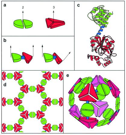
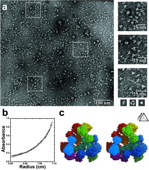
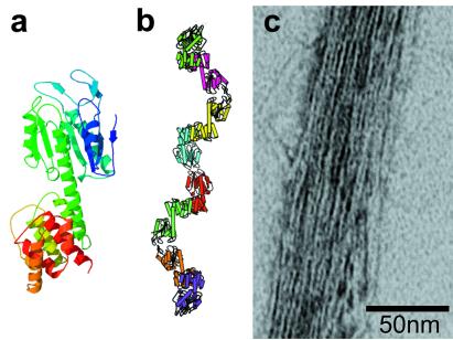
A general strategy is described for designing proteins that self assemble into large symmetrical nanomaterials, including molecular cages, filaments, layers, and porous materials. In this strategy, one molecule of protein A, which naturally forms a self-assembling oligomer, A(n), is fused rigidly to one molecule of protein B, which forms another self-assembling oligomer, B(m). The result is a fusion protein, A-B, which self assembles with other identical copies of itself into a designed nanohedral particle or material, (A-B)(p). The strategy is demonstrated through the design, production, and characterization of two fusion proteins: a 49-kDa protein designed to assemble into a cage approximately 15 nm across, and a 44-kDa protein designed to assemble into long filaments approximately 4 nm wide. The strategy opens a way to create a wide variety of potentially useful protein-based materials, some of which share similar features with natural biological assemblies.


Figures

Figure 1
Figure 1
A general strategy for designing fusion proteins that assemble into symmetric nanostructures. (a) The green semicircle represents a natural dimeric protein (i.e., a protein that associates with one other copy of itself), whereas the red shape represents a trimeric protein. The symmetry axes of the natural oligomers are shown. (b) The two natural proteins are combined by genetic methods into a single fusion protein. Each of the original natural proteins serves as an “oligomerization domain” in the designed fusion protein. Two different hypothetical fusion proteins are shown to illustrate that the oligomerization domains can be joined rigidly in different geometries. (c) A ribbon diagram of a fusion protein showing one method for joining two oligomerization domains (red and green) in a relatively rigid fashion. One of the natural oligomerization domains must end in an α-helical conformation, and the other must begin in an α-helical conformation. The two are then linked by a short stretch of amino acids (blue) that have a strong tendency to adopt an α-helical conformation. Thus, the two oligomerization domains are joined physically in a predictable orientation. (d) A designed fusion protein self assembles into a particular kind of nanostructure that depends on the geometry of the symmetry axes belonging to its component oligomerization domains (Table 1). A molecular layer arises from an arrangement like that in b (Left). (e) A cubic cage arises from an arrangement like the one in b (Right).
Figure 2
Figure 2
Characterization of a designed tetrahedral protein cage. (a) Negatively stained electron micrographs show images of discrete particles. The images left in the heavy-atom stain are consistent with the sizes of the largest faces of the cage. For size comparison (shown to scale, Bottom Right), three simulated images were calculated from the atomic coordinates of the cage in three orientations where it would make the most extensive contacts with the surface of the electromagnetic support grid. As a rough approximation, it was assumed that the complex would leave a footprint in a layer of heavy-atom stain 15 Å thick. (b) Equilibrium sedimentation shows that the major component has a molecular mass of approximately 550 kDa, corresponding roughly to 11.3 subunits (close to the anticipated value of 12). A small degree of polymorphism is evident from the residual difference between the experimental and theoretical curves. (c) A stereo model of the tetrahedral protein cage as it was intended to assemble from 12 copies of the 49-kDa engineered fusion protein (shown in Fig. 1c). The view is through one of the four large openings in the cage. The particle radius is approximately 9 nm, and the edge length is approximately 15 nm. The separate protein subunits are colored differently.
Figure 3
Figure 3
Electron microscopy and model of a designed protein filament. (a) A ribbon model of a single molecule of the designed fusion protein. (b) A ribbon model of the protein filament as it was intended to assemble, with separate protein molecules colored differently. (c) Negatively stained electron micrograph of a bundle of filaments formed by the designed fusion protein. The bundle is 15–20 filaments across and reveals details indicative of the individual dimeric oligomerization domains that make up the fusion protein. In addition to bundles, networks of filaments were also observed in other micrographs.

References

    1. Whitesides G M, Mathias J P, Seto C T. Science. 1991;254:1312–1319. - PubMed
    1. Herron N, Thorn D L. Adv Mater. 1998;10:1173–1184.
    1. Kroto H W, Heath J R, O'Brien S C, Curl R F, Smalley R E. Nature (London) 1985;318:162–163.
    1. Zhang Y, Seeman N C. J Am Chem Soc. 1994;116:1661–1669.
    1. Huang H Y, Remsen E E, Kowalewski T, Wooley K L. J Am Chem Soc. 1999;121:3805–3806.

Publication types

LinkOut - more resources