. 2001 Feb 27;98(5):2284-8.
doi: 10.1073/pnas.031430998. Epub 2001 Feb 20.

The human mitochondrial deoxynucleotide carrier and its role in the toxicity of nucleoside antivirals

Affiliations

The human mitochondrial deoxynucleotide carrier and its role in the toxicity of nucleoside antivirals

V Dolce et al. Proc Natl Acad Sci U S A. .

Abstract

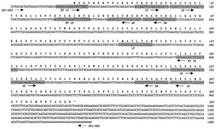
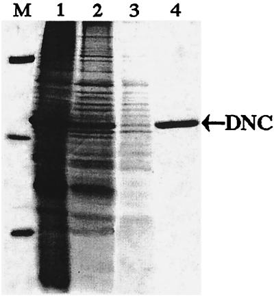
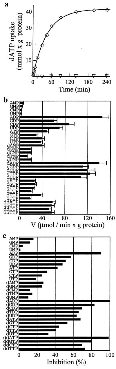
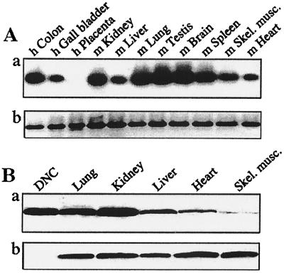
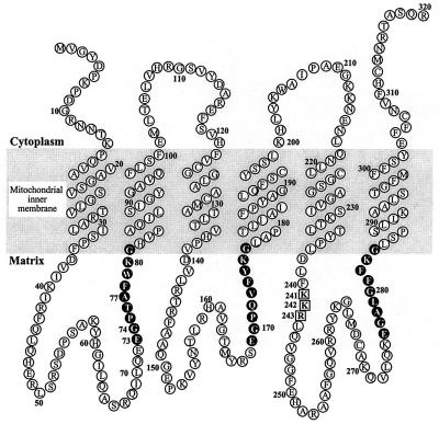
The synthesis of DNA in mitochondria requires the uptake of deoxynucleotides into the matrix of the organelle. We have characterized a human cDNA encoding a member of the family of mitochondrial carriers. The protein has been overexpressed in bacteria and reconstituted into phospholipid vesicles where it catalyzed the transport of all four deoxy (d) NDPs, and, less efficiently, the corresponding dNTPs, in exchange for dNDPs, ADP, or ATP. It did not transport dNMPs, NMPs, deoxynucleosides, nucleosides, purines, or pyrimidines. The physiological role of this deoxynucleotide carrier is probably to supply deoxynucleotides to the mitochondrial matrix for conversion to triphosphates and incorporation into mitochondrial DNA. The protein is expressed in all human tissues that were examined except for placenta, in accord with such a central role. The deoxynucleotide carrier also transports dideoxynucleotides efficiently. It is likely to be medically important by providing the means of uptake into mitochondria of nucleoside analogs, leading to the mitochondrial impairment that underlies the toxic side effects of such drugs in the treatment of viral illnesses, including AIDS, and in cancer therapy.


Figures

Figure 1
Figure 1
Sequence of a human cDNA and the encoded DNC. Amino acids are numbered from 1–320. An asterisk denotes the stop codon. Primers and probes are shaded. The nested primers 1F/2F and 1R/2R and probe 1P were used to confirm the EST sequence. The partial cDNA sequence was extended in 3′ and 5′ directions with primers AP1 and AP2 and nested oligonucleotides 3F/4F or 3R/4R, respectively. Primers RT 1F and RT 1R and probe RT 1P were used in reverse transcription–PCR experiments. Horizontal arrows pointing right and left indicate that primers were synthesized as shown or as the complement, respectively.
Figure 2
Figure 2
Purification of DNC by Ni+-agarose affinity chromatography. Proteins were separated by SDS/PAGE and stained with Coomassie blue. Lane M, markers (BSA, carbonic anhydrase, and cytochrome c); lane 1, sarkosyl extract of inclusion bodies; lane 2, pH 6.8 eluate; lane 3, pH 6.5 eluate; lane 4, purified DNC, eluted at pH 6.2. The position of DNC is indicated on the right by an arrow.
Figure 3
Figure 3
Time course of dATP/ADP exchange and substrate specificity of human DNC. (a) Time course of [α-35S]dATP/ADP exchange in proteoliposomes reconstituted with the recombinant DNC. [α-35S]dATP (1 mM) was added to proteoliposomes containing 10 mM ADP (⋄) or 10 mM NaCl (▿) (b) Dependence of DNC activity on internal substrate. Proteoliposomes were preloaded internally with various substrates (concentration 10 mM). Transport was started by addition of 20 μM [α-35S]dATP and stopped after 2 min. The values are means ± SD of at least three experiments. (c) Inhibition of the rate of [α-35S]dATP uptake by external substrates. Proteoliposomes were preloaded internally with 10 mM ADP. Transport was started by adding 125 μM [α-35S]dATP and stopped after 2 min. External substrates (concentration 0.5 mM) were added together with [α-35S]dATP. The extents of inhibition (%) from a representative experiment are reported. The control value for uninhibited exchange was 0.45 mmol/min per gram of protein.
Figure 4
Figure 4
Expression of human DNC in various tissues. Analysis of total RNA from human (h) and mouse (m) tissues (A). (a) Hybridization of cDNA fragments for the DNC with probe RT 1P. (b) Ethidium-bromide-stained cDNA fragments for β-actin. (B) Immunodetection of the DNC in mitochondria isolated from rat tissues. In a and b, mitochondria (150 μg of protein) and human DNC (75 ng) were exposed to antisera to the DNC and subunit IV of the cytochrome c oxidase, respectively.
Figure 5
Figure 5
Folding of the DNC in the inner membranes of mitochondria. The topography of the six transmembrane α-helices is based on the hydrophobic profile of the sequence in Fig. 1. Each of the three tandem repeats in the sequence is folded into two transmembrane α-helices with a large intervening hydrophilic loop. The three repetitive elements are linked by shorter loops. The cytoplasmic and matrix locations of the various features are based on experimental evidence of locations of analogous features in other members of the family of mitochondrial carriers. The sequences in black are related to the DNA-binding domain of the nuclear receptor family. Residues 241–243 (in squares) correspond to the sequence RRR at residues 234–236 of the ANC from Saccharomyces cerevisiae.

References

    1. Walker J E. Curr Opin Struct Biol. 1992;2:519–526.
    1. Palmieri F. FEBS Lett. 1994;346:48–54. - PubMed
    1. Palmieri F, van Ommen B. In: Frontiers in Cellular Bioenergetics. Papa S, Guerrieri F, Tager J M, editors. New York: Kluwer Academic/Plenum; 1999. pp. 489–519.
    1. Palmieri L, De Marco V, Iacobazzi V, Palmieri F, Runswick M J, Walker J E. FEBS Lett. 1997;410:447–451. - PubMed
    1. Palmieri L, Lasorsa F M, De Palma A, Palmieri F, Runswick M J, Walker J E. FEBS Lett. 1997;417:114–118. - PubMed

Publication types

Associated data