. 2001 Feb 27;98(5):2358-63.
doi: 10.1073/pnas.051629298. Epub 2001 Feb 20.

Transverse relaxation-optimized NMR spectroscopy with the outer membrane protein OmpX in dihexanoyl phosphatidylcholine micelles

Affiliations

Transverse relaxation-optimized NMR spectroscopy with the outer membrane protein OmpX in dihexanoyl phosphatidylcholine micelles

C Fernández et al. Proc Natl Acad Sci U S A. .

Abstract

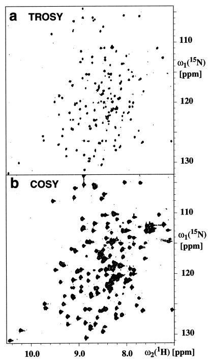
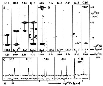
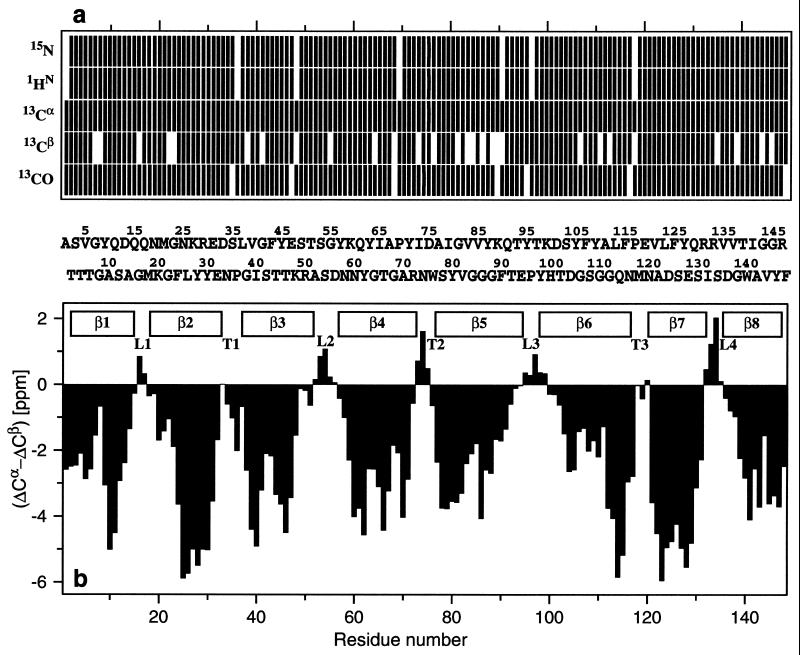
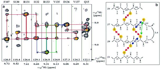
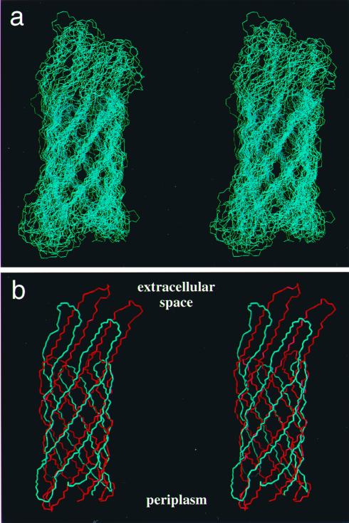
The (2)H,(13)C,(15)N-labeled, 148-residue integral membrane protein OmpX from Escherichia coli was reconstituted with dihexanoyl phosphatidylcholine (DHPC) in mixed micelles of molecular mass of about 60 kDa. Transverse relaxation-optimized spectroscopy (TROSY)-type triple resonance NMR experiments and TROSY-type nuclear Overhauser enhancement spectra were recorded in 2 mM aqueous solutions of these mixed micelles at pH 6.8 and 30 degrees C. Complete sequence-specific NMR assignments for the polypeptide backbone thus have been obtained. The (13)C chemical shifts and the nuclear Overhauser effect data then resulted in the identification of the regular secondary structure elements of OmpX/DHPC in solution and in the collection of an input of conformational constraints for the computation of the global fold of the protein. The same type of polypeptide backbone fold is observed in the presently determined solution structure and the previously reported crystal structure of OmpX determined in the presence of the detergent n-octyltetraoxyethylene. Further structure refinement will have to rely on the additional resonance assignment of partially or fully protonated amino acid side chains, but the present data already demonstrate that relaxation-optimized NMR techniques open novel avenues for studies of structure and function of integral membrane proteins.


Figures

Figure 1
Figure 1
Contour plots of 15N—1H correlation spectra measured with a 2 mM solution of 2H,13C,15N-labeled OmpX in DHPC micelles (20 mM phosphate/100 mM NaCl/0.05% NaN3/300 mM DHPC/3% 2H2O/solvent H2O, pH 6.8, T = 30°C) at a 1H resonance frequency of 750 MHz. (a) 2D [15N,1H]-TROSY. (b) 2D [15N,1H]-COSY. The two spectra were recorded and processed identically (see text). The measuring time for each spectrum was 4 h.
Figure 2
Figure 2
(a) [ω2(13C), ω3(1H)] strips from a 3D [15N,1H]-TROSY-HNCA spectrum. The strips were taken at the 15N chemical shifts (indicated at the bottom of the strips) of residues 12–16 and are centered about the corresponding 1HN chemical shifts. At the top of each strip, the sequence-specific assignment is indicated by the one-letter amino acid symbol and the sequence position. Horizontal and vertical lines connect the intraresidual and sequential HNCA connectivities and, thus, outline the sequential assignment pathway. (b) Same presentation as a for the corresponding 3D HNCA spectrum. (c) Cross sections along the ω2(13C) dimension through the peaks in a and b. The upper traces correspond to the 3D [15N,1H]-TROSY-HNCA spectrum shown in a, and the lower ones correspond to the conventional 3D HNCA spectrum shown in b.
Figure 3
Figure 3
(a) Survey of the NMR assignments for OmpX/DHPC obtained by TROSY-type triple-resonance experiments. The residues for which the 1HN, 15N, 13Cα, 13Cβ, and 13CO chemical shifts have been assigned are indicated by vertical bars in the respective rows. In the center, separating a and b, the amino acid sequence is indicated by the one-letter amino acid symbols, where the entries have been distributed over two rows, i.e., residue 1 is in the upper row, residue 2 is in the lower row, etc. (b) Plot of (ΔCα − ΔCβ) vs. the amino acid sequence. ΔCα and ΔCβ were obtained as the differences between the experimental 13Cα and 13Cβ chemical shifts in OmpX/DHPC and the corresponding random coil shifts. The value of (ΔCα − ΔCβ) for a particular residue i represents the average over the three consecutive residues i − 1, i and i + 1, and was calculated as follows: formula image (41). Negative values of (ΔCα − ΔCβ) indicate that residue i is located in a regular β-strand (positive values would indicate location in a regular helical structure). The positions of the regular secondary structure elements in the crystal structure of OmpX are indicated at the top, and the external loops (L) and periplasmatic turns (T) are labeled according to Vogt and Schulz (23).
Figure 4
Figure 4
(a) [ω1(1H), ω3(1H)] strips from a 3D [1H,1H]-NOESY-[15N,1H]-TROSY spectrum taken at the 15N chemical shifts of the residues indicated at the top of each strip. Yellow dots indicate diagonal peaks, blue lines connect sequential dNN cross-peaks with the diagonal peaks, green lines indicate medium-range dNN(i,i+2) NOEs, and red lines show long-range dNN(i,j) NOEs between neighboring β-strands. (b) Positions in the OmpX structure of the NOEs in the spectral regions shown in a. The same color code as in a is used to identify the amide protons that correspond to the diagonal peaks in a and for the arrows that indicate the NOE connectivities.
Figure 5
Figure 5
Stereoviews of the polypeptide backbone fold in OmpX. (a) Superposition of the 20 dyana conformers that were selected to represent the NMR structure of OmpX. The superposition is for pairwise global best fit of the N, Cα, and C′ backbone atoms of the β-sheet amino acid residues in conformers 2–20 with the corresponding atoms in the conformer with the smallest residual target function value (Table 1). (b) Comparison of the mean NMR structure (blue) and the x-ray crystal structure (red) after superposition as in a. Periplasmatic and extracellular spaces are indicated according to ref. . The figure was prepared with the program molmol (43).

References

    1. Walker J E, Saraste M. Curr Opin Struct Biol. 1996;6:457–459. - PubMed
    1. Kühlbrandt W, Gouaux E. Curr Opin Struct Biol. 1999;9:445–447. - PubMed
    1. Bowie J. Curr Opin Struct Biol. 2000;10:435–437. - PubMed
    1. Wüthrich K. NMR of Proteins and Nucleic Acids. New York: Wiley; 1986.
    1. Cavanagh J, Fairbrother W J, Palmer A G, III, Skelton N J. Protein NMR Spectroscopy. Principles and Practice. New York: Academic; 1996.

Publication types

MeSH terms

LinkOut - more resources