. 2001 Feb 27;98(5):2381-6.
doi: 10.1073/pnas.041603398.

Presenilin-1 binds cytoplasmic epithelial cadherin, inhibits cadherin/p120 association, and regulates stability and function of the cadherin/catenin adhesion complex

Affiliations

Presenilin-1 binds cytoplasmic epithelial cadherin, inhibits cadherin/p120 association, and regulates stability and function of the cadherin/catenin adhesion complex

L Baki et al. Proc Natl Acad Sci U S A. .

Abstract

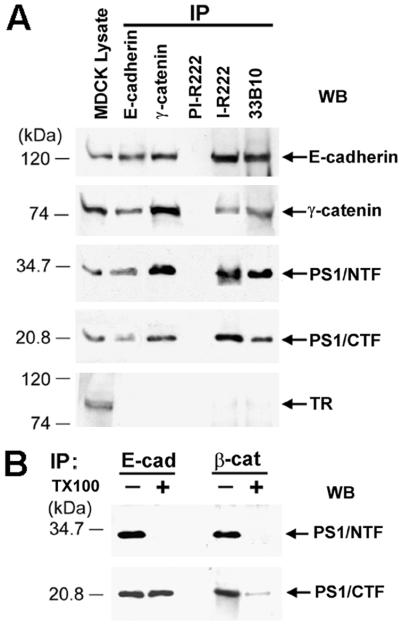
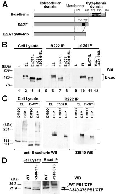
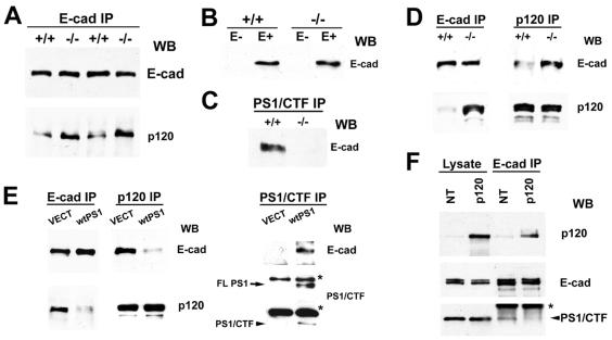
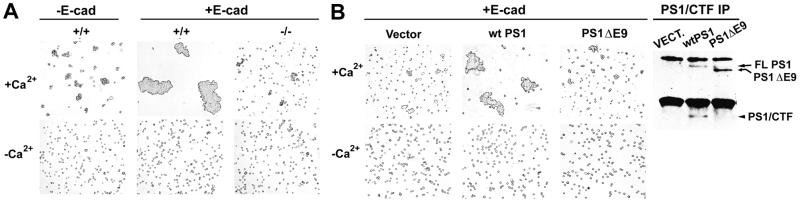
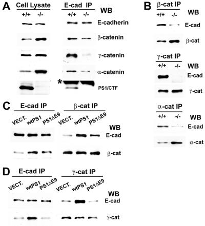
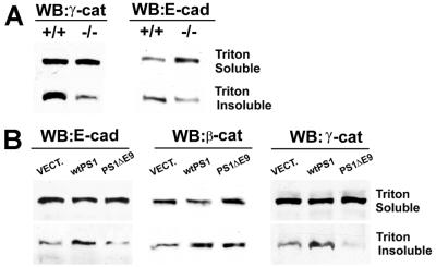
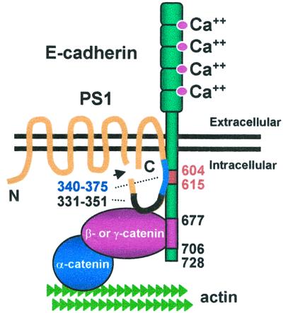
Here we show that presenilin-1 (PS1), a protein involved in Alzheimer's disease, binds directly to epithelial cadherin (E-cadherin). This binding is mediated by the large cytoplasmic loop of PS1 and requires the membrane-proximal cytoplasmic sequence 604-615 of mature E-cadherin. This sequence is also required for E-cadherin binding of protein p120, a known regulator of cadherin-mediated cell adhesion. Using wild-type and PS1 knockout cells, we found that increasing PS1 levels suppresses p120/E-cadherin binding, and increasing p120 levels suppresses PS1/E-cadherin binding. Thus PS1 and p120 bind to and mutually compete for cellular E-cadherin. Furthermore, PS1 stimulates E-cadherin binding to beta- and gamma-catenin, promotes cytoskeletal association of the cadherin/catenin complexes, and increases Ca(2+)-dependent cell-cell aggregation. Remarkably, PS1 familial Alzheimer disease mutant DeltaE9 increased neither the levels of cadherin/catenin complexes nor cell aggregation, suggesting that this familial Alzheimer disease mutation interferes with cadherin-based cell-cell adhesion. These data identify PS1 as an E-cadherin-binding protein and a regulator of E-cadherin function in vivo.


Figures

Figure 1
Figure 1
(A) PS1 forms complexes with E-cadherin and γ-catenin. Extract of confluent MDCK cells in TNE buffer plus 1% digitonin (9) was treated with the antibodies indicated at the top of the figure, and the resulting IPs were probed on WBs with antibodies against the proteins indicated on the right. For reference, 20 μg of MDCK cell lysate was also probed. TR, transferrin receptor; PI-R222, rabbit 222 preimmune serum; I-R222, rabbit 222 antiserum; 33B10, anti-PS1/CTF monoclonal antibody. (B) Triton X-100 effects on PS1/β-catenin and PS1/E-cadherin complexes. Extracts of confluent MDCK cells in 1% digitonin were immunoprecipitated with antibodies against E-cadherin (E-cad) or β-catenin (β-cat), and the resulting IPs were washed in the presence (+) or absence (−) of 1% Triton X-100 (TX100). The remaining immunocomplexes were collected by centrifugation and probed on WBs with antibody R222 (PS1/NTF) or 33B10 (PS1/CTF).
Figure 2
Figure 2
PS1 binds E-cadherin independently of β- or γ-catenin. Cytoplasmic residues 604–615 are required for PS1/E-cadherin binding. (A) Schematic representation of mouse E-cadherin and E-cadherin deletion constructs EΔC71 and EΔC71Δ604–615, which is a derivative of EΔC71 and lacks cytoplasmic residues 604–615 (see text). Constructs were prepared as previously described (25). (B) PS1 binding to E-cadherin constructs. Extracts (1% digitonin) from L cells (L, lanes 1, 5, and 9) or L cells transfected with mouse E-cadherin (EL, lanes 2, 6, and 10), construct EΔC71 (EΔC71L, lanes 3, 7, and 11), or construct EΔC71Δ604–615 (EΔC71Δ604–615L, lanes 4, 8, and 12) were treated with antibody R222 (lanes 5–8) or anti-p120 antibody (lanes 9–12), and the resulting IPs were probed on WBs with anti-E-cadherin antibody H108. For reference, 20 μg of cell lysates was probed (lanes 1–4). (C) Confluent EL or EΔC71L cells were incubated either with the crosslinking agent dithiobis (succinimidylpropionate) (DSP) in DMSO or with DMSO alone, and cell extracts were then prepared in RIPA buffer in the presence of SDS to inhibit noncovalent associations. Lysates were treated with antibody R222 (R222 IP), and the resulting IPs were probed on WBs with anti-E-cadherin (H108) or anti-PS1/CTF (33B10) antibodies. Twenty micrograms of cell lysate was probed with H108 antibody. (D) PS1 construct Δ340–375 does not bind E-cadherin. HEK293 cells expressing WT PS1 (WT) or a PS1 construct with amino acid deletion 340–375 (Δ340–375) were lysed in TNE plus 1% digitonin. Lysates were treated with anti-E-cadherin antibody (E-cad IP), and the resulting IPs were probed on WBs with antibody 33B10. Twenty micrograms of cell lysates was also probed.
Figure 3
Figure 3
PS1 inhibits p120 binding to E-cadherin. (A) E-cadherin IPs (E-cad IP) in TNE plus 1% Triton X-100 from either PS1−/− mouse embryos (−/−) or their PS1+/+ littermates (+/+) were probed with anti-E-cadherin (E-cad) or anti-p120 (p120) antibodies. (B) Extracts (1% SDS) of confluent nontransfected (E−) or E-cadherin-transfected (E+) PS1+/+ (+/+) and PS1−/− (−/−) mouse fibroblasts were probed with anti-E-cadherin antibody. (C) Extracts (1% Triton X-100) of confluent E-cadherin-expressing PS1+/+ (+/+) or PS1−/− (−/−) fibroblasts were immunoprecipitated with antibody 33B10 (PS1/CTF IP), and the resulting IPs were probed with anti-E-cadherin (E-cad) antibody. (D) Extracts (1% Triton X-100) of E-cadherin-transfected PS1 +/+ (+/+) or PS1−/− (−/−) fibroblasts were immunoprecipitated with anti-E-cadherin (E-cad IP) or anti-p120 (p120 IP) antibodies, and the resulting IPs were probed with anti-E-cadherin (E-cad) or anti-p120 (p120) antibodies. (E) E-cadherin-transfected PS1−/− fibroblasts were transiently transfected either with vector (VECT) or with WT PS1 (wtPS1), and Triton X-100 extracts were immunoprecipitated with anti-E-cadherin (E-cad IP), anti-p120 (p120 IP), or anti-PS1/CTF (PS1/CTF IP) antibodies. The resulting IPs were probed with anti-E-cadherin (E-cad), anti-p120 (p120), or anti-PS1/CTF antibodies. FLPS1, full-length PS1. The asterisks indicate IgGs. (F) Extracts (1% Triton X-100) of untransfected (NT) EL cells or EL cells transiently transfected with p120 (p120) were treated with anti-E-cadherin antibodies (E-cad IP), and the resulting IPs were probed with antibodies against the proteins indicated on the right. The asterisk indicates IgGs.
Figure 4
Figure 4
PS1 stimulates Ca2+-dependent cell–cell aggregation. (A) Nontransfected (−E-cad) PS1 +/+ (+/+) or E-cadherin-transfected (+E-cad) PS1 +/+ (+/+) and PS1−/− (−/−) fibroblasts were allowed to aggregate for 4 h in the presence (+Ca2+) or absence (−Ca2+) of calcium (see Materials and Methods). (B) E-cadherin-expressing PS1−/− fibroblasts, transiently transfected with vector (Vector), WT PS1 (wtPS1), or PS1 FAD mutant ΔE9 (PS1ΔE9), were assayed for aggregation in the presence or absence of calcium. (Right) Extracts (1% Triton X-100) from the transiently transfected cultures shown in B were treated with antibody 33B10, and the resulting IPs (PS1/CTF IP) were analyzed for expression of the transfected PS1 proteins.
Figure 5
Figure 5
PS1 stabilizes the cadherin/catenin complex. (A) Extracts of confluent E-cadherin-transfected PS1+/+ and PS1−/− fibroblasts in 1% Triton X-100 were treated with anti-E-cadherin antibody, and the resulting IPs (E-cad IP, Right), along with total cell lysate in 1% SDS (Left), were probed on WBs with antibodies against the proteins indicated at the right of the figure. The asterisk denotes low-molecular-weight IgGs. (B) Anti-β-catenin, anti-γ-catenin, or anti-α-catenin IPs (Top, Middle, and Bottom, respectively) prepared from E-cadherin-expressing PS1+/+ and PS1−/− fibroblasts were probed with anti-E-cadherin (E-cad), anti-β-catenin (β-cat), anti-γ-catenin (γ-cat), or anti-α-catenin (α-cat) antibodies. (C and D) Extracts (1% Triton X-100) of E-cadherin-expressing PS1−/− fibroblasts transiently transfected with vector (VECT), WT PS1 (wtPS1), or PS1 FAD mutant ΔE9 (PS1ΔE9) were treated with anti-E-cadherin (E-cad IP), anti-β-catenin (β-cat IP) (C), or anti-γ-catenin (γ-cat IP) (D) antibodies, and the resulting IPs were probed with antibodies against the proteins indicated at the right of the figure. E-cad, E-cadherin; β-cat, β-catenin; γ-cat, γ-catenin.
Figure 6
Figure 6
PS1 promotes cytoskeletal association of the cadherin/catenin complexes. (A) Confluent cultures of E-cadherin-transfected PS1+/+ or PS1−/− fibroblasts were extracted in 1% Triton X-100, and insoluble fractions were solubilized in urea buffer (see Materials and Methods). Sixty micrograms of protein of the Triton X-100-soluble fraction or 75 μg of the insoluble fraction was then probed with antibodies against γ-catenin (γ-cat) or E-cadherin (E-cad). (B) Extracts from E-cadherin-transfected PS1−/− fibroblasts, transiently transfected with vector (VECT), WT PS1 (wtPS1), or PS1 FAD mutant ΔE9 (PS1ΔE9), were prepared and probed as in A, plus anti-β-catenin (β-cat) antibodies.
Figure 7
Figure 7
Schematic representation of hypothesized interactions between E-cadherin, β- οr γ-catenin, and PS1. β-Catenin or γ-catenin binds E-cadherin at amino acids 677–706. PS1 C-terminal fragment binds E-cadherin close to the membrane at amino acids 604–615 (required for PS1/E-cadherin complex formation). For more details see text.

References

    1. Sherrington R, Rogaev E I, Liang Y, Rogaeva E A, Levesque G, Ikeda M, Chi H, Lin C, Li G, Holman K, et al. Nature (London) 1995;375:754–760. - PubMed
    1. Elder G A, Tezapsidis N, Carter J, Shioi J, Bouras C, Li H C, Johnston J M, Efthimiopoulos S, Friedrich V L, Jr, Robakis N K. J Neurosci Res. 1996;45:308–320. - PubMed
    1. Podlisny M B, Citron M, Amarante P, Sherrington R, Xia W, Zhang J, Diehl T, Levesque G, Fraser P, Haass C, et al. Neurobiol Dis. 1997;3:325–337. - PubMed
    1. Thinakaran G, Borchelt D R, Lee M K, Slunt H H, Spitzer L, Kim G, Ratovitsky T, Davenport F, Nordstedt C, Seeger M, et al. Neuron. 1996;17:181–190. - PubMed
    1. De Strooper B, Annaert W, Cupers P, Saftig P, Craessaerts K, Mumm J S, Schroeter E H, Schrijvers V, Wolfe M S, Ray W J, et al. Nature (London) 1999;398:518–522. - PubMed

Publication types

LinkOut - more resources