. 2001 Feb 27;98(5):2387-92.
doi: 10.1073/pnas.041606398.

An isolated, surface-expressed I domain of the integrin alphaLbeta2 is sufficient for strong adhesive function when locked in the open conformation with a disulfide bond

Affiliations

An isolated, surface-expressed I domain of the integrin alphaLbeta2 is sufficient for strong adhesive function when locked in the open conformation with a disulfide bond

C Lu et al. Proc Natl Acad Sci U S A. .

Abstract

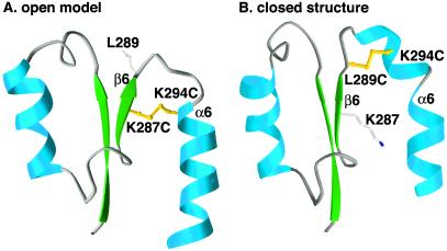
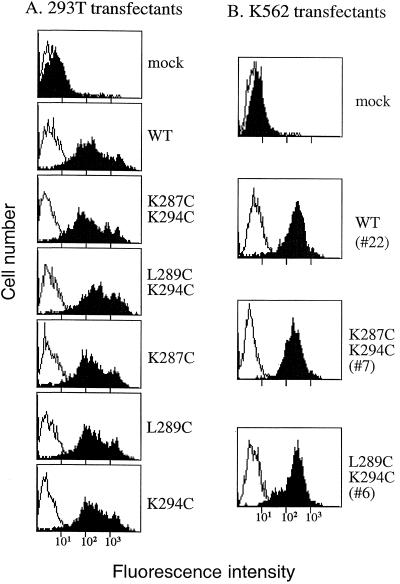
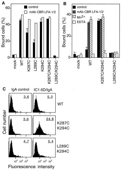
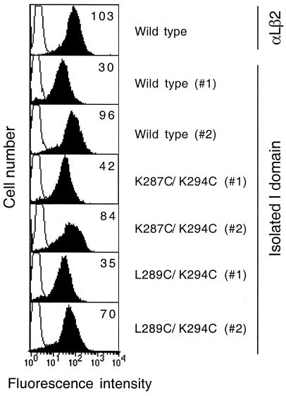
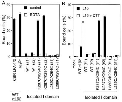
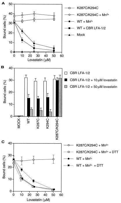
We introduced disulfide bonds to lock the integrin alphaLbeta2 I domain in predicted open, ligand binding or closed, nonbinding conformations. Transfectants expressing alphaLbeta2 heterodimers containing locked-open but not locked-closed or wild-type I domains constitutively adhered to intercellular adhesion molecule-1 (ICAM-1) substrates. Locking the I domain closed abolished constitutive and activatable adhesion. The isolated locked-open I domain bound as well as the activated alphaLbeta2 heterodimer, and binding was abolished by reduction of the disulfide. Lovastatin, which binds under the conformationally mobile C-terminal alpha-helix of the I domain, inhibited binding to ICAM-1 by alphaLbeta2 with wild-type, but not locked-open I domains. These data establish the importance of conformational change in the alphaL I domain for adhesive function and show that this domain is sufficient for full adhesive activity.


Figures

Figure 1
Figure 1
Predicted disulfide bonds that are selective for open or closed conformers of the αL I domain. The K287C/K294C mutation is modeled in the open conformer (A), and the L289C/K294C mutation is modeled in the closed conformer (B). For clarity, ribbon diagrams show only residues 254 to 305 of the I domain. The models were superimposed by using residues not involved in conformational shifts and are shown in exactly the same orientation. The downward shift in the α6 helix in A compared with B is readily apparent. The remodeling of the loop connecting β6 and α6 is accompanied by a reversal in the orientation of the sidechain of residue 289. Sidechains for residues 287, 289, and 294 are shown as ball and stick. Prepared with ribbons.**
Figure 2
Figure 2
Cell surface expression of αLβ2 cysteine substitution mutants. (A) 293T transient transfectants. (B) K562 stable transfectants. Wild-type or mutant αL cDNA was cotransfected with β2 cDNA in 293T cells or K562 cells. Cell surface expression of the αLβ2 complex was determined by immunofluorescent flow cytometry of the transfectants with mAb TS2/4 (shaded histogram) specific for αL in the αLβ2 complex, or the nonbinding IgG X63 (open histogram). Numbers in parentheses are clone numbers of the K562 stable transfectants.
Figure 3
Figure 3
Ligand binding activity of αLβ2 cysteine substitution mutants. (A) Binding of 293T transient transfectants to immobilized ICAM-1. (B) Binding of K562 stable transfectants to ICAM-1. Binding of the transfectants to immobilized ICAM-1 was determined in L15/FBS that contains Ca2+ and Mg2+ in the presence of the activating mAb CBR LFA-1/2 (10 μg/ml) or the control nonbinding IgG X63 (control), or in the absence of Ca2+ and Mg2+ and presence of 1 mM Mn2+ or 5 mM EDTA. Results are mean ± SD of triplicate samples and are representative of at least three experiments; some error bars are too small to be visible. (C) Binding of soluble ICAM-1-IgA Fc fusion protein (IC1–5D/IgA) to K562 stable transfectants. K562 transfectants were incubated with 100 μg/ml IC1–5D/IgA (Right) or control human IgA (Left), followed by incubation with FITC-labeled anti-human IgA and flow cytometry. Numbers on the upper right corner of each histogram plot are mean fluorescent intensity. Results are representative of three experiments.
Figure 4
Figure 4
Cell surface expression of the isolated αL I domains. The wild-type αL I domain, the open mutant K287C/K294C I domain, and the closed mutant L289C/K294C I domain were expressed on the surface of K562 transfectants with the PDGFR transmembrane domain. The level of cell surface expression was determined by immunofluorescent flow cytometry using mAb TS1/22 to the I domain (filled histograms). Binding of the control X63 IgG is shown as open histograms. The mean fluorescent intensity of TS1/22 binding is shown in the upper right corner of each plot. Two individual clones (#1 and #2) from each I domain transfectant are shown.
Figure 5
Figure 5
Ligand binding activity of the isolated αL I domains. (A) Comparison of binding to immobilized ICAM-1 of activated wild-type αLβ2 and isolated, mutant αL I domains expressed in K562 transfectants. Binding of K562 transfectants expressing isolated I domains was in the presence of Ca2+ and Mg2+ (control), or in absence of Ca2+ and Mg2+ and presence of 1 mM EDTA. Binding of K562 transfectants expressing wild-type αLβ2 was in the presence of Ca2+ and Mg2+ and mAb CBR LFA-1/2 (10 μg/ml), or in absence of Ca2+ and Mg2+ and presence of 1 mM Mn2+. (B) Effect of disulfide reduction by DTT on binding of K562 transfectants to immobilized ICAM-1. Binding was performed in L15/FBS in the presence or absence of 10 mM DTT.
Figure 6
Figure 6
Lovastatin, a small molecule inhibitor of LFA-1, inhibits the function of wild-type αLβ2 but not of αLβ2 with a mutant, open I domain. (A and C) K562 transfectants. (B) 293T transfectants. Cells expressing wild-type αLβ2 or αLβ2 with mutant I domains, or mock-transfected cells, were preincubated with different concentrations of lovastatin at 37°C for 15 min before addition to ICAM-1-coated 96-well plastic plates. In C, cells were incubated in the presence or absence of 2 mM DTT at 37°C for 15 min before lovastatin treatment. Results are expressed as mean ± SD of three independent experiments in duplicate.

References

    1. Larson R S, Springer T A. Immunol Rev. 1990;114:181–217. - PubMed
    1. Springer T A. Cell. 1994;76:301–314. - PubMed
    1. Gahmberg C G, Tolvanen M, Kotovuori P. Eur J Biochem. 1997;245:215–232. - PubMed
    1. Diamond M S, Springer T A. Curr Biol. 1994;4:506–517. - PubMed
    1. Humphries M J. Biochem Soc Trans. 2000;28:311–339. - PubMed

Publication types

MeSH terms

LinkOut - more resources