. 2001 Feb 27;98(5):2443-8.
doi: 10.1073/pnas.041493198. Epub 2001 Feb 20.

IL-4 inhibits osteoclast formation through a direct action on osteoclast precursors via peroxisome proliferator-activated receptor gamma 1

Affiliations

IL-4 inhibits osteoclast formation through a direct action on osteoclast precursors via peroxisome proliferator-activated receptor gamma 1

A C Bendixen et al. Proc Natl Acad Sci U S A. .

Abstract

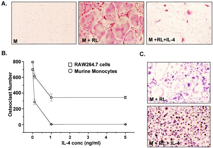
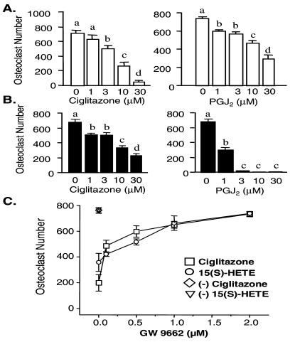
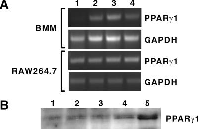
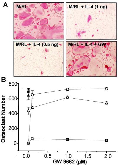
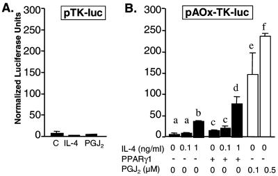
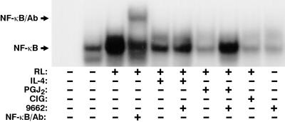
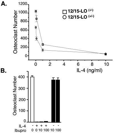
IL-4 is a pleiotropic immune cytokine secreted by activated T(H)2 cells that inhibits bone resorption both in vitro and in vivo. The cellular targets of IL-4 action as well as its intracellular mechanism of action remain to be determined. We show here that IL-4 inhibits receptor activator of NF-kappaB ligand-induced osteoclast differentiation through an action on osteoclast precursors that is independent of stromal cells. Interestingly, this inhibitory effect can be mimicked by both natural as well as synthetic peroxisome proliferator-activated receptor gamma1 (PPARgamma1) ligands and can be blocked by the irreversible PPARgamma antagonist GW 9662. These findings suggest that the actions of IL-4 on osteoclast differentiation are mediated by PPARgamma1, an interpretation strengthened by the observation that IL-4 can activate a PPARgamma1-sensitive luciferase reporter gene in RAW264.7 cells. We also show that inhibitors of enzymes such as 12/15-lipoxygenase and the cyclooxygenases that produce known PPARgamma1 ligands do not abrogate the IL-4 effect. These findings, together with the observation that bone marrow cells from 12/15-lipoxygenase-deficient mice retain sensitivity to IL-4, suggest that the cytokine may induce novel PPARgamma1 ligands. Our results reveal that PPARgamma1 plays an important role in the suppression of osteoclast formation by IL-4 and may explain the beneficial effects of the thiazolidinedione class of PPARgamma1 ligands on bone loss in diabetic patients.


Figures

Figure 1
Figure 1
IL-4 suppresses M-CSF/RANKL-induced osteoclast formation in both murine BMs and RAW264.7 cells. (A) BMs were treated with M-CSF (M) (10 ng/ml) or M-CSF plus RANKL (RL) (30 ng/ml) in the absence or presence of IL-4 (1 ng/ml) for 10 days, stained for TRAP, and photographed at ×20. (B) BMs (1 × 10⁵ cells per well) and RAW264.7 cells (2 × 10³ cells per well) were plated in triplicate and induced with M-CSF/RANKL in the absence or presence of increasing amounts of IL-4. Multinucleated (more than three nuclei), TRAP-positive osteoclasts were quantitated after 10 days (BMs) or 5 days (RAW264.7). Mean ± SE, n = 3. (C) IL-4 treatment suppresses osteoclastogenesis and results in enhanced macrophage formation in BMs. BMs were treated with the indicated factors for 10 days and then fixed and stained for α-naphthylacetate esterase.
Figure 2
Figure 2
Ciglitazone and 15d-PGJ2 act via PPARγ to suppress M-CSF/RANKL-induced osteoclast formation in primary murine myeloid (BMs) and RAW264.7 cells. (A) BMs were plated in triplicate at 105 cells per well and treated with M-CSF/RANKL in the presence of vehicle (ethanol or DMSO), ciglitazone (1–30 μM), or 15d-PGJ2 (PGJ2) (1–30 μM) for a period of 7–10 days, and then multinucleated (more than three), TRAP-positive osteoclasts were quantitated. (B) RAW264.7 cells were plated in triplicate at 2 × 10³ cells per well and treated as in A. Multinucleated, TRAP-positive osteoclasts were quantitated after 5 days. (C) The PPARγ1 antagonist GW 9662 prevents ciglitazone- and 15(S)-HETE-induced suppression of osteoclast formation in BMs. Cells were incubated with M-CSF/RANKL and either vehicle, ciglitazone (30 μM) or 15(S)-HETE (30 μM), in the presence of increasing concentrations of GW 9662. Mean ± SE, n = 3 (b, c, and d are significant vs. a at P < 0.05).
Figure 3
Figure 3
Detection of PPARγ in BM and RAW264.7 cells. (A) Regulation of PPARγ1 mRNA by GM-CSF, M-CSF, and IL-4 in BMs but not in RAW264.7 cells. BMs and RAW264.7 cells were cultured in the absence or presence of one of the following cytokines for 24 h: vehicle (lane 1); mGM-CSF (10 ng/ml, lane 2), M-CSF (10 ng/ml, lane 3), or IL-4 (10 ng/ml, lane 4). Total RNA was isolated, treated with DNase, and subjected to reverse transcription–PCR analysis. (B) Detection of PPARγ1 protein. BMs were incubated untreated (lane 1) or were treated with 10 ng/ml M-CSF for 48 h (lane 2) or 72 h (lane 3) or with 100 ng/ml M-CSF for 48 h (lane 4) or 72 h (lane 5), and nuclear extracts (75 μg) were evaluated by Western analysis.
Figure 4
Figure 4
IL-4 suppresses M-CSF/RANKL-induced osteoclast formation in BMs. (A) BMs were treated with M-CSF (10 ng/ml) and RANKL (30 ng/ml) in the absence or presence of IL-4 (1 ng/ml) for 10 days, stained for TRAP, and photographed at ×20. (Upper) Cells that have been cultured in the absence (Left) or presence (Right) of 1 ng/ml IL-4. (Lower) Cells that have been cultured in the presence of 0.5 ng/ml IL-4 in the absence (Left) or presence (Right) of the PPARγ1 antagonist GW 9662 (2 μM). (B) BMs (1 × 10⁵ cells per well) were plated in triplicate and induced with M-CSF/RANKL in the absence (●, ■, ▴) or presence of IL-4 (□, 1 ng/ml; ○, 0.5 ng/ml; ▵, 0.1 ng/ml) and increasing amounts of GW 9662 (0, 0.1, 1 and 2 μM). Multinucleated (more than three nuclei), TRAP-positive osteoclasts were quantitated after 10 days. Mean ± SE, n = 3.
Figure 5
Figure 5
IL-4 induces activation of PPARγ1-mediated transcription in RAW264.7 cells. (A) RAW264.7 cells were transfected with pTK-luc and stimulated with either vehicle, IL-4 (1 ng/ml) or 15d-PGJ2 (PGJ2) (0.5 μM) in the presence of 0.5% serum. (B) RAW264.7 cells were transfected with pAOx-TK-luc without or with pCMX-PPARγ1 (10 ng) and stimulated with IL-4 or 15d-PGJ2 (PGJ2) as indicated. Mean ± SE, n = 3 (b, e, and f are significant vs. a, and d is significant vs. c at P < 0.05).
Figure 6
Figure 6
IL-4 and PPARγ ligands suppress RANKL-dependent activation of NF-κB. RAW264.7 cells (2.5 × 10⁶ cells per plate) were pretreated for 30 min with IL-4 (1 ng/ml), 15d-PGJ2 (PGJ2) (0.5 μM), ciglitazone (CIG) (10 μM), and/or GW 9662 (1 μM) as indicated and then treated for 30 min with RANKL (100 ng/ml). Electrophoretic mobility-shift assay was carried out as indicated in Materials and Methods. Lane 1 represents the free probe. The NF-κB specific DNA complexes and supershifted NF-κB are indicated by the arrows.
Figure 7
Figure 7
12/15-LO and COX-1/COX-2 are not required for IL-4 action. (A) BMs were isolated from mice heterozygous (+/−) or homozygous (−/−) for the 12/15-lipoxygenase null allele, plated in triplicate (1 × 10⁵ cells per well), and induced with M-CSF/RANKL in the absence or presence of increasing amounts of IL-4 (0.1, 1, and 10 ng/ml). Multinucleated (more than three nuclei), TRAP-positive osteoclasts were quantitated after 10 days. (B) BMs from normal mice were treated for 10 days with M-CSF/RANKL, IL-4 (0.5 ng/ml), or ibuprofen (10 or 100 μM) as indicated. Mean ± SE, n = 3.

References

    1. Manolagas S C, Jilka R L. N Engl J Med. 1995;332:305–311. - PubMed
    1. Turner R T, Riggs B L, Spelsberg T C. Endocr Rev. 1994;15:275–300. - PubMed
    1. Marcus R, Feldman D, Kelsey J, editors. Osteoporosis. San Diego: Academic; 1996.
    1. Roodman G D. Exp Hematol. 1999;27:1229–1241. - PubMed
    1. Suda T, Takahashi N, Udagawa N, Jimi E, Gillespie M T, Martin T J. Endocr Rev. 1999;20:345–357. - PubMed

Publication types

MeSH terms

LinkOut - more resources