. 2001 Feb 27;98(5):2461-6.
doi: 10.1073/pnas.041449398. Epub 2001 Feb 13.

Production of zebrafish germ-line chimeras from embryo cell cultures

Affiliations

Production of zebrafish germ-line chimeras from embryo cell cultures

C Ma et al. Proc Natl Acad Sci U S A. .

Abstract

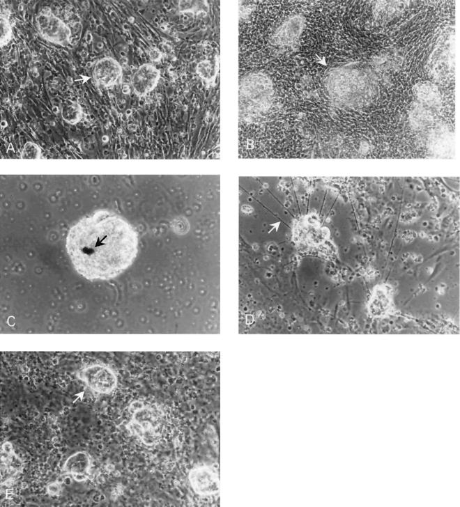
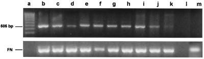
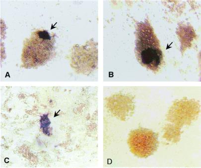
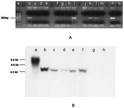
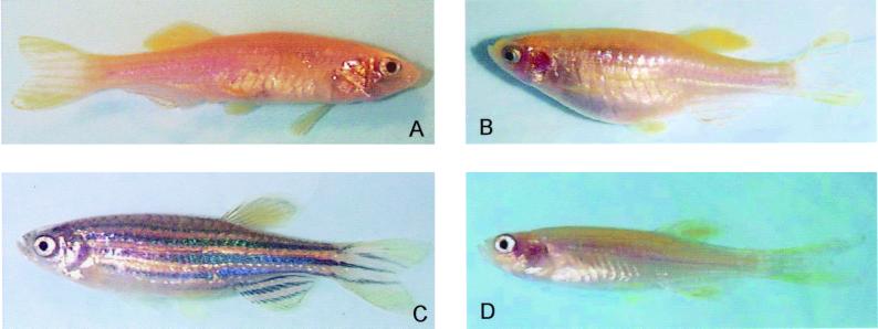
Although the zebrafish possesses many characteristics that make it a valuable model for genetic studies of vertebrate development, one deficiency of this model system is the absence of methods for cell-mediated gene transfer and targeted gene inactivation. In mice, embryonic stem cell cultures are routinely used for gene transfer and provide the advantage of in vitro selection for rare events such as homologous recombination and targeted mutation. Transgenic animals possessing a mutated copy of the targeted gene are generated when the selected cells contribute to the germ line of a chimeric embryo. Although zebrafish embryo cell cultures that exhibit characteristics of embryonic stem cells have been described, successful contribution of the cells to the germ-cell lineage of a host embryo has not been reported. In this study, we demonstrate that short-term zebrafish embryo cell cultures maintained in the presence of cells from a rainbow trout spleen cell line (RTS34st) are able to produce germ-line chimeras when introduced into a host embryo. Messenger RNA encoding the primordial germ-cell marker, vasa, was present for more than 30 days in embryo cells cocultured with RTS34st cells or their conditioned medium and disappeared by 5 days in the absence of the spleen cells. The RTS34st cells also inhibited melanocyte and neuronal cell differentiation in the embryo cell cultures. These results suggest that the RTS34st splenic-stromal cell line will be a valuable tool in the development of a cell-based gene transfer approach to targeted gene inactivation in zebrafish.


Figures

Figure 1
Figure 1
Phase contrast photomicrographs of zebrafish embryo cell cultures. (a) Culture (24 h) of embryo cell aggregates (arrow) on a feeder layer of RTS34st cells. (b) Culture (20 day old) of embryo cell aggregates (arrow) grown on an RTS34st feeder layer. (c) Culture (15 day old) maintained in the absence of feeder cells showing the presence of melanocytes (arrow). (d) Culture (10 day old) maintained in the absence of feeder cells showing the presence of neurites (arrow), indicating that neural cell differentiation has occurred. The neurites begin to appear in the culture on approximately day 5. (e) Culture (25 day old) of embryo cells in RTS34st cell-conditioned medium without a feeder layer, illustrating embryo cell aggregates (arrow) on a monolayer of embryo fibroblasts.
Figure 2
Figure 2
Reverse transcription–PCR analysis of vasa mRNA in zebrafish embryo cell cultures. cDNA was synthesized from total RNA obtained from embryo cell cultures. PCR amplification was performed with vasa-specific primers designed to generate a 505-bp product. Product identity was confirmed by sequencing. Lane a, MW markers; lanes b, c, and d, embryo cells maintained for 5, 15, and 25 days, respectively, in RTS34st cell-conditioned medium; lanes e, f, and g, embryo cells maintained for 5, 15, and 25 days, respectively, on an RTS34st feeder layer; lane h, embryo cells maintained at first for 24 days in RTS34st cell-conditioned medium, and then (after passaging) for 8 days on an RTS34st feeder layer; lanes i, j, and k, embryo cell cultures maintained for 1, 3, and 5 days, respectively, in the absence of RTS34st feeder cells or cell-conditioned medium; lane l, negative control (no template); lane m, RTS34st cells cultured in the absence of zebrafish embryo cells. Primers that amplify fibronectin cDNA were used to control for equal amounts of sample in each lane.
Figure 3
Figure 3
Distribution of vasa-positive embryo cells. Cultures maintained for 3 days (a) and 8 days (b) in RTS34st cell-conditioned medium or 3 days on RTS34st feeder cells (c) were examined by in situ hybridization by using a vasa-specific antisense probe. The control culture (d) was grown for 8 days in conditioned medium and hybridized with sense probe. [Magnification = ×200 (a, b, and d), and = ×100 (c).]
Figure 4
Figure 4
PCR (A) and Southern blot analysis (B) of genomic DNA showing the presence of neo sequences. (A) Genomic DNA isolated from individual F1 fish (lanes b–m) produced from a single spawning of GASSI fish that were injected as embryos with cultured cells and bred with noninjected GASSI individuals. DNA was amplified with neo-specific primers designed to generate a 392-bp product. Product identity was confirmed by sequencing. neo sequences were detected in lanes b, i, k, and l. Lanes a, n, and o are molecular weight markers, positive control (neo-containing plasmid template) and negative control (no template), respectively. (B) Southern blot analysis of genomic DNA by using a neo-specific probe. The same integration pattern for neo sequences was observed in DNA isolated from B7–43 fish (lane b), cell cultures derived from B7–43 embryos (lane c), individual F1 fish (lanes d and e) obtained from a GASSI chimera that was injected at the blastula stage with cultured B7–43 embryo cells and bred with a noninjected GASSI fish, and an F2 individual (lane f) obtained by breeding positive F1 siblings. Lane a, DNA isolated from fish embryo cells transfected in culture with neo-containing plasmid showing a different integration pattern. Lanes g and h, DNA isolated from a GASSI fish and another nontransgenic line of zebrafish.
Figure 5
Figure 5
Zebrafish phenotypes. (a) A chimeric zebrafish from a GASSI embryo that had been injected at the blastula stage with cultured cells derived from B7–43 embryos. Melanocyte pigmentation is absent on the body of the chimera. (b) GASSI fish that was bred with the chimera shown in a to produce (c) F1 individuals that exhibited a pigmentation pattern characteristic of B7–43 and (d) the nonpigmented GASSI phenotype.

References

    1. Streisinger G, Walker C, Dower N, Knauber D, Singer F. Nature (London) 1981;291:293–296. - PubMed
    1. Nusslein-Volhard C. Science. 1994;266:572–574. - PubMed
    1. Driever W, Stemple D, Schier A, Solnica-Krezel L. Trends Genet. 1994;10:152–159. - PubMed
    1. Mullins M C, Nusslein-Volhard C. Curr Opin Genet Dev. 1993;3:648–654. - PubMed
    1. van Eeden F J M, Granato M, Odenthal J, Haffter P. In: Methods in Cell Biology. Detrich H W, Westerfield M, Zon L I, editors. San Diego: Academic; 1999. pp. 21–41. - PubMed

Publication types

LinkOut - more resources