. 2001 Feb 27;98(5):2532-7.
doi: 10.1073/pnas.051609898. Epub 2001 Feb 13.

Somatic mosaicism in Fanconi anemia: evidence of genotypic reversion in lymphohematopoietic stem cells

Affiliations

Somatic mosaicism in Fanconi anemia: evidence of genotypic reversion in lymphohematopoietic stem cells

J J Gregory Jr et al. Proc Natl Acad Sci U S A. .

Abstract

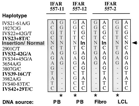
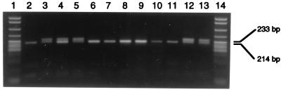
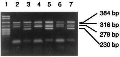
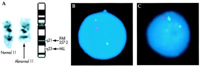
Somatic mosaicism has been observed previously in the lymphocyte population of patients with Fanconi anemia (FA). To identify the cellular origin of the genotypic reversion, we examined each lymphohematopoietic and stromal cell lineage in an FA patient with a 2815-2816ins19 mutation in FANCA and known lymphocyte somatic mosaicism. DNA extracted from individually plucked peripheral blood T cell colonies and marrow colony-forming unit granulocyte-macrophage and burst-forming unit erythroid cells revealed absence of the maternal FANCA exon 29 mutation in 74.0%, 80.3%, and 86.2% of colonies, respectively. These data, together with the absence of the FANCA exon 29 mutation in Epstein-Barr virus-transformed B cells and its presence in fibroblasts, indicate that genotypic reversion, most likely because of back mutation, originated in a lymphohematopoietic stem cell and not solely in a lymphocyte population. Contrary to a predicted increase in marrow cellularity resulting from reversion in a hematopoietic stem cell, pancytopenia was progressive. Additional evaluations revealed a partial deletion of 11q in 3 of 20 bone marrow metaphase cells. By using interphase fluorescence in situ hybridization with an MLL gene probe mapped to band 11q23 to identify colony-forming unit granulocyte-macrophage and burst-forming unit erythroid cells with the 11q deletion, the abnormal clone was exclusive to colonies with the FANCA exon 29 mutation. Thus, we demonstrate the spontaneous genotypic reversion in a lymphohematopoietic stem cell. The subsequent development of a clonal cytogenetic abnormality in nonrevertant cells suggests that ex vivo correction of hematopoietic stem cells by gene transfer may not be sufficient for providing life-long stable hematopoiesis in patients with FA.


Figures

Figure 1
Figure 1
Haplotypes for SNPs segregating in family IFAR 557. Both common and rare polymorphisms were typed on genomic DNA extracted from PB from the father (IFAR 557–11) and the mother (IFAR 557–12) and on DNA extracted from the LCL and fibroblasts from the patient (IFAR 557–2). N, normal allele; In, 2815–2816ins19; bold type, common polymorphisms; regular type, rare polymorphisms; *, rare haplotype. Phase is deduced from data from other subjects, indicating that the rare SNPs are found only as a single haplotype with the common variants as shown.
Figure 2
Figure 2
Mutation screening of individually plucked hematopoietic precursors. FANCA exon 29 was PCR-amplified from individually plucked hematopoietic precursors. Presence of the normal allele is indicated by a 214-bp product, and the presence of the 2815–2816ins19 mutation is indicated by a 233-bp product. Lanes: 1 and 14, pBR322 MspI markers; 2, IFAR 557–11 PB; 3, IFAR 557–12 PB; 4, IFAR 557–2 PB; 5–9, IFAR 557–2 BFU-E; 10–12, IFAR 557–2 CFU-GM; 13, IFAR 557–12 BFU-E.
Figure 3
Figure 3
FANCA polymorphism screening in flow-sorted colonies. Paired multiplex allele-specific PCRs for each allele of three FANCA polymorphisms were performed on a BFU-E colony positive for 2815–2816ins19 (lanes 2 and 3), PB (lanes 4 and 5), and a BFU-E colony negative for 2815–2816ins19 (lanes 6 and 7). Reaction 1 (lanes 2, 4, and 6) results in PCR products in the presence of the IVS23+8T (279 bp), IVS32–42G (316 bp), and IVS42+29T (230 bp) alleles. Reaction 2 (lanes 3, 5, and 7) results in PCR products in the presence of the IVS23+8C, IVS32–42A, and IVS42+29C alleles. Homozygosity for a polymorphism for a given sample is indicated by the presence of a band in one reaction but not the other, whereas heterozygosity for a given sample is indicated by the presence of a band in both reactions. A 384-bp control product from exon 6 of FANCA is present in both reactions. Lane 1, pBR322 MspI markers.
Figure 4
Figure 4
(A) G banded chromosome 11 homologs from IFAR 557–2 bone marrow sample. (Left) Normal chromosome 11. (Center) Abnormal chromosome 11 resulting in loss of material distal to the breakpoint in band 11q21. (Right) Idiogram showing normal G banding pattern of chromosome 11 at approximately 400-band-level resolution and the position of the MLL locus. (B) Image of a normal interphase cell from an IFAR 557–2 colony. FISH shows two chromosome 11 centromere signals (green) and two MLL gene signals (red). (C) Image of a cell harboring the abnormal chromosome 11 from an IFAR 557–2 colony. FISH shows two chromosome 11 centromere signals (green), but only one MLL signal (red).

References

    1. Auerbach A D, Buchwald M, Joenje H. In: The Metabolic and Molecular Basis of Inherited Diseases. 8th Ed. Scriver C R, Beadet A L, Sly W S, Valle D, Childs B, Vogelstein B, editors. New York: McGraw–Hill; 2001. pp. 753–768.
    1. Joenje H, Levitus M, Waisfisz Q, D'Andrea A, Garcia-Higuera I, Pearson T, van Berkel C G, Rooimans M A, Morgan N, Mathew C G, et al. Am J Hum Genet. 2000;67:759–762. - PMC - PubMed
    1. Lo Ten Foe J R, Rooimans M A, Bosnoyan-Collins L, Alon N, Wijker M, Parker L, Lightfoot J, Carreau M, Callen D F, Savoia A, et al. Nat Genet. 1996;14:320–323. - PubMed
    1. Fanconi Anemia/Breast Cancer Consortium. Nat Genet. 1996;14:324–328.
    1. Strathdee C A, Duncan A M, Buchwald M. Nat Genet. 1992;1:196–198. - PubMed

Publication types