Comparative Study
. 2001 Feb 27;98(5):2740-5.
doi: 10.1073/pnas.051612298. Epub 2001 Feb 13.

Comparative genomics of the restriction-modification systems in Helicobacter pylori

Affiliations
Comparative Study

Comparative genomics of the restriction-modification systems in Helicobacter pylori

L F Lin et al. Proc Natl Acad Sci U S A. .

Abstract

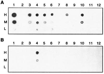
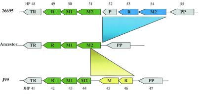
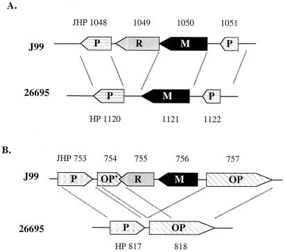
Helicobacter pylori is a Gram-negative bacterial pathogen with a small genome of 1.64-1.67 Mb. More than 20 putative DNA restriction-modification (R-M) systems, comprising more than 4% of the total genome, have been identified in the two completely sequenced H. pylori strains, 26695 and J99, based on sequence similarities. In this study, we have investigated the biochemical activities of 14 Type II R-M systems in H. pylori 26695. Less than 30% of the Type II R-M systems in 26695 are fully functional, similar to the results obtained from strain J99. Although nearly 90% of the R-M genes are shared by the two H. pylori strains, different sets of these R-M genes are functionally active in each strain. Interestingly, all strain-specific R-M genes are active, whereas most shared genes are inactive. This agrees with the notion that strain-specific genes have been acquired more recently through horizontal transfer from other bacteria and selected for function. Thus, they are less likely to be impaired by random mutations. Our results also show that H. pylori has extremely diversified R-M systems in different strains, and that the diversity may be maintained by constantly acquiring new R-M systems and by inactivating and deleting the old ones.


Figures

Figure 1
Figure 1
Antibody assay to measure methyltransferase activities by using rabbit primary antibodies raised specifically against m6A or m4C. Dot blot assays were performed by spotting total DNAs, isolated from clones expressing individual H. pylori methylase genes, onto a nitrocellulose BA85 membrane (18). Positive signals were detected by using a secondary anti-rabbit antibody conjugated with horseradish peroxidase (18). Lanes 1–12 represent HP1351–1352; pHKUV5 vector (a negative control); HP91–92; HP1366–1367-1368; HP54; HP50–51; HP263; HP478; HP481; HP910; HP1471–1472; and HP1121, respectively. (A) MTase dot blot assay using antibodies against m6A. (B) MTase dot blot assay using antibodies against m4C. Three dilutions of DNA samples were spotted: H (high), 0.45 μg; M (medium), 0.15 μg; and L (low), 0.05 μg.
Figure 2
Figure 2
Schematic diagram showing the possible evolution of HP49–54 in strain 26695 and JHP42–46 in strain J99 from a putative ancestral locus. The shared Type II R-M systems are shown in green. The strain-specific Type II R-M system in strain 26695 is shown in blue, and the strain-specific Type II R-M system in J99 is shown in yellow. Functional genes are shown in solid colors and inactive genes are patterned with white dots. Non-R-M genes are shown in gray. R, restriction endonuclease gene; M, DNA methylase gene; P, putative gene; TR, transcriptional regulator gene; PP, proline permease gene. HP numbers are marked above the genes and JHP numbers are marked below the genes.
Figure 3
Figure 3
Determination of the recognition specificities of H. pylori endonucleases by double digestion. DNA endonucleotic cleavage activity of isoschizomers of MboI and MboII, encoded by HP91–92 and HP1366–1367-1368, respectively. (A) Bacteriophage lambda DNA was digested with MboI (lane 1), MboI and purified endonuclease HpyAIII (lane 2), or HpyAIII (lane 3). (B) Lambda DNA was digested with MboII (lane 1), MboII and purified endonuclease HpyAII (lane 2), or HpyAII alone (lane 3).
Figure 4
Figure 4
Schematic diagram showing the alignment of homologous loci. (A) The alignment of homologous loci where the strain-specific R-M system genes of JHP755–756 are inserted in strain J99. (B) The deletion of an endonuclease gene (homologue of JHP1049) in strain 26695. R, restriction endonuclease gene (shown in gray); M, DNA methylase gene (shown in black). Non-R-M genes are shown in patterned boxes. P, putative gene; OP, osmoprotection protein gene; OP', 5′ end of a truncated OP gene. JHP numbers are marked above the genes and HP numbers are marked below the genes.
Figure 5
Figure 5
Schematic diagram showing the dynamic acquisition and loss of R-M system genes. Different R-M systems are shown in different colors. Triangles, R gene; circles, M gene. Orphan genes are not connected to each other. Functional genes are shown in solid colors and inactive genes are patterned with white dots.

References

    1. Blaser M J. Sci Am. 1996;274(2):104–107. - PubMed
    1. Covacci A, Telford J L, Del Giudice G, Parsonnet J, Rappuoli R. Science. 1999;284:1328–1333. - PubMed
    1. Wang G, Humayun M Z, Taylor D E. Trends Microbiol. 1999;7:488–493. - PubMed
    1. Tomb J F, White O, Kerlavage A R, Clayton R A, Sutton G G, Fleischmann R D, Ketchum K A, Klenk H P, Gill S, Dougherty B A, et al. Nature (London) 1997;388:539–547. - PubMed
    1. Alm R A, Ling L S, Moir D T, King B L, Brown E D, Doig P C, Smith D R, Noonan B, Guild B C, deJonge B L, et al. Nature (London) 1999;397:176–180. - PubMed

Publication types

Substances