. 2001 Feb 27;98(5):2764-9.
doi: 10.1073/pnas.051628498. Epub 2001 Feb 13.

Synergistic contributions of cyclin-dependant kinase 5/p35 and Reelin/Dab1 to the positioning of cortical neurons in the developing mouse brain

Affiliations

Synergistic contributions of cyclin-dependant kinase 5/p35 and Reelin/Dab1 to the positioning of cortical neurons in the developing mouse brain

T Ohshima et al. Proc Natl Acad Sci U S A. .

Abstract

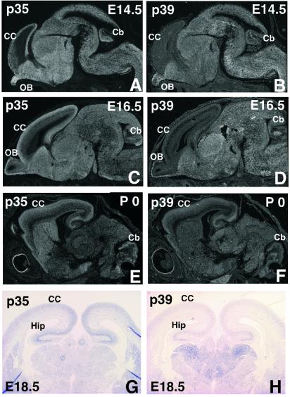
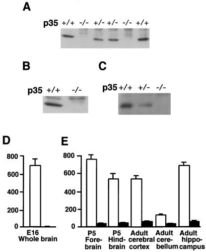
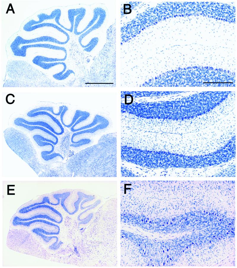
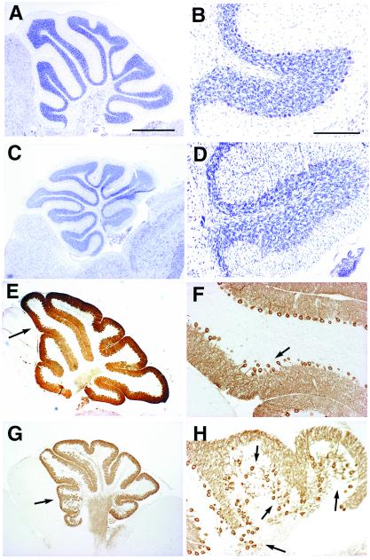
Cyclin-dependent kinase (Cdk) 5 is a unique member of the Cdk family, because Cdk5 kinase activity is detected only in the nervous tissue. Two neuron-specific activating subunits of Cdk5, p35 and p39, have been identified. Overlapping expression pattern of these isoforms in the embryonic mouse brain and the significant residual Cdk5 kinase activity in brain homogenate of the p35-/- mice indicate the redundant functions of the Cdk5 activators in vivo. Severe neuronal migration defects in p35-/-Cdk5 +/- mice further support the idea that the redundant expression of the Cdk5 activators may cause a milder phenotype in p35-/- mice compared with Cdk5-/- mice. Mutant mice lacking either Cdk5 or p35 exhibit certain similarities with Reelin/Dab1-mutant mice in the disorganization of cortical laminar structure in the brain. To elucidate the relationship between Cdk5/p35 and Reelin/Dab1 signaling, we generated mouse lines that have combined defects of these genes. The addition of heterozygosity of either Dab1 or Reelin mutation to p35-/- causes the extensive migration defects of cortical neurons in the cerebellum. In the double-null mice of p35 and either Dab1 or Reelin, additional migration defects occur in the Purkinje cells in the cerebellum and in the pyramidal neurons in the hippocampus. These additional defects in neuronal migration in mice lacking both Cdk5/p35 and Reelin/Dab1 indicate that Cdk5/p35 may contribute synergistically to the positioning of the cortical neurons in the developing mouse brain.


Figures

Figure 1
Figure 1
Overlapping expression of Cdk5 activating subunits. Comparison of p35 and p39 gene expression in sagittal brain sections from E14.5 and E16.5 embryos and newborns with 35S-labeled riboprobes (AF) and in coronal sections from E18.5 embryos with digoxigenin-labeled probes (G and H) by in situ hybridization. Expressions of p35 and p39 overlap in the brain except in the cerebral cortex at E14.5 and E16.5. CC, cerebral cortex; Cb, cerebellum; OB, olfactory bulb; Hip, hippocampus.
Figure 2
Figure 2
p35−/− mice retain residual Cdk5 kinase activity. (AC) Western blot analysis of p35 protein of the brain homogenate (A and B) and immunoprecipitate with anti-Cdk5 antibody (C) from the indicated genotype at postnatal day (P) 2 by using p35 antibodies that recognize the carboxyl terminus of p35 protein (A and C) and the N terminus of p35 protein (B). No p35 protein was detected in p35−/− brain, and a reduced amount of p35 protein was detected in the p35+/− brain (A and C). (D and E) Cdk5 kinase activity in brain homogenates from Cdk5+/+ (white bar) and Cdk5−/− (D) and p35+/+ (white bar) and p35−/− (black bar) homogenates (E). The diagram represents mean ± standard deviation (n = 3) of Cdk5 activity. Cdk5 kinase activity is expressed as pmol of phosphate incorporated per hr per mg protein. Approximately 10 and 20% residual Cdk5 kinase activity is detected in the cerebral cortex and cerebellum of p35−/− brain, respectively, but none in the Cdk5−/− brain.
Figure 3
Figure 3
Defects in neuronal migration in p35−/− mice are accentuated by Cdk5 heterozygosity. Parasagittal sections of cerebella of wild-type littermates (A and B) and p35−/− (C and D) at P21, and p35−/−Cdk5+/− at P35 (E and F) are stained with toluidine blue. B, D, and F are higher magnification of A, C, and E, respectively. In the p35−/− cerebellum, the alignment of Purkinje cells is disturbed slightly in some areas, and significant numbers of granule cells are found in the molecular layer (D). In the cerebellum of p35−/−Cdk5+/− mice, defects in the alignment of Purkinje cells are accentuated in the most areas (E and F). Purkinje cells are found occasionally in the granule cell layer, and increased numbers of granule cells are trapped in the molecular layer (F). (Bar in A, 1 mm; bar in B, 200 μm.)
Figure 4
Figure 4
Migration defects of cerebellar cortical neurons in p35−/− mice are accentuated by Dab1 heterozygosity. Comparative histology of cerebella of Dab1yot/+ (A and B), p35−/− (E and F), and p35−/−Dab1yot/+ (C, D, G, and H) at P21 in the parasagittal sections stained with toluidine blue (AD) and with anti-IP3R antibody (EH). B, D, F, and H are higher magnifications of A, C, E, and G, respectively. Arrows in E and G indicate magnified areas in F and H, respectively. Subtle disturbances of the alignment of Purkinje cells are observed in p35−/− (arrows in F). Extensive migration defects of Purkinje cells and granule cells are observed in p35−/−Dab1yot/+ compared with p35−/− cerebellum (D, G, and H). (Bar in A, 1 mm. A, C, E, and G are identical magnification. Bar in B, 200 μm.)
Figure 5
Figure 5
Double-null mutant mice for p35 and Reelin/Dab1 display additional defects. (AF) Comparison of the histological appearance of cerebella between Dab1yot/yot (A, C, and E) and p35−/−Dab1yot/yot at P22 (B, D, and F) in parasagittal sections stained with toluidine blue (AD) and with anti-calbindin antibody (E and F). The molecular layer is defective in the p35−/−Dab1yot/yot cerebellum (B, D, and F). The arrow in F indicates the molecular layer with dendrites of Purkinje cells. (Bar in B and E, 500 μm; bar in D, 200 μm.) Mo, molecular layer; Gr, granule cell layer. (GM) Coronal sections of Nissl staining at the level of hippocampus (GJ) and of septum (KM) from wild type (K), p35−/− (G and L), and Dab1yot/yot (H) at P21 and p35−/−Dab1yot/yot (M) and p35−/−rl/rl (I and J) at P15. Accentuated disorganization of the pyramidal cell layer is observed in p35−/−rl/rl (arrows in I and J). The upper part of the septum is hypoplastic in p35−/− (arrow in L). Separation of the upper part of the septum is observed in p35−/−Dab1yot/yot mice (arrow in M). (Bar in G, 400 μm; bar in J, 80 μm; bar in K, 200 μm.)

Similar articles

Cited by

References

    1. Ohshima T, Ward J M, Huh C-G, Longnecker G, Veeranna, Pant H C, Brady R O, Martin L J, Kulkarni A B. Proc Natl Acad Sci USA. 1996;93:11173–11178. - PMC - PubMed
    1. Chae T, Kwon Y T, Bronson R, Dikkes P, Li E, Tsai L-H. Neuron. 1997;18:29–42. - PubMed
    1. Hellmich M R, Pant H C, Wada E, Betty J F. Proc Natl Acad Sci USA. 1992;89:10867–10871. - PMC - PubMed
    1. Lew J, Winkfein R J, Paudel H K, Wang J H. J Biol Chem. 1992;267:25922–25926. - PubMed
    1. Meyerson M, Enders G H, Wu C L, Su L K, Gorka C, Nelson C, Harlow E, Tsai L-H. EMBO J. 1992;11:2909–2917. - PMC - PubMed

Publication types

MeSH terms