

Morphological abnormalities in the brains of estrogen receptor beta knockout mice
- PMID: 11226319
- PMCID: PMC30218
- DOI: 10.1073/pnas.041617498
Morphological abnormalities in the brains of estrogen receptor beta knockout mice
Abstract
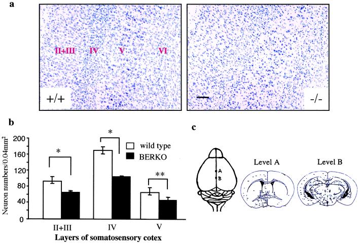
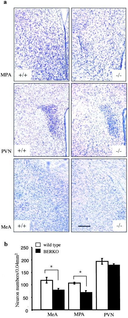
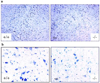
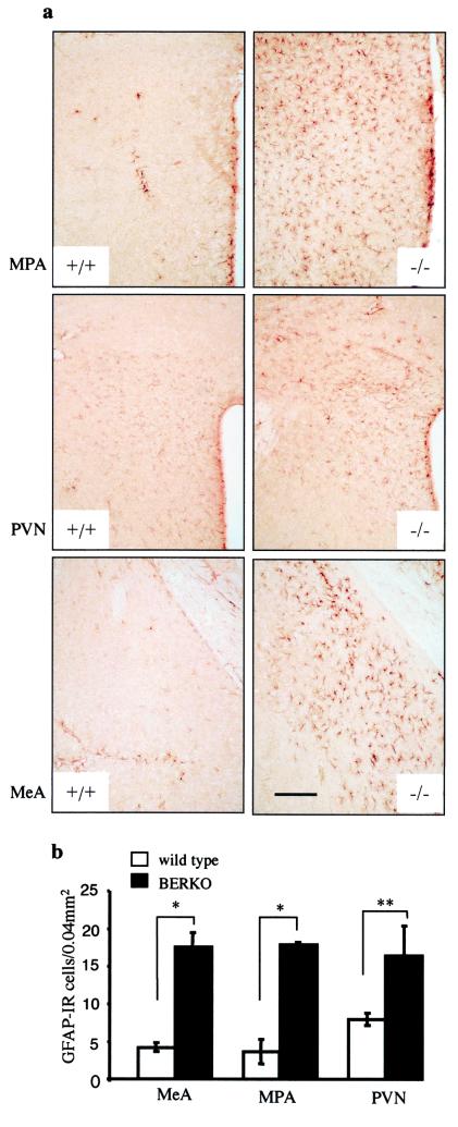
Estrogen receptor beta (ERbeta) is expressed at high levels in both neurons and glial cells of the central nervous system. The development of ERbeta knockout (BERKO) mice has provided a model to study the function of this nuclear receptor in the brain. We have found that the brains of BERKO mice show several morphological abnormalities. There is a regional neuronal hypocellularity in the brain, with a severe neuronal deficit in the somatosensory cortex, especially layers II, III, IV, and V, and a remarkable proliferation of astroglial cells in the limbic system but not in the cortex. These abnormalities are evident as early as 2 mo of age in BERKO mice. As BERKO mice age, the neuronal deficit becomes more pronounced, and, by 2 yr of age, there is degeneration of neuronal cell bodies throughout the brain. This is particularly evident in the substantia nigra. We conclude that ERbeta is necessary for neuronal survival and speculate that this gene could have an important influence on the development of degenerative diseases of the central nervous system, such as Alzheimer's disease and Parkinson's disease, as well as those resulting from trauma and stroke in the brain.
Figures
Similar articles
-
Expression of estrogen receptors (alpha, beta) and androgen receptor in serotonin neurons of the rat and mouse dorsal raphe nuclei; sex and species differences.Neurosci Res. 2004 Jun;49(2):185-96. doi: 10.1016/j.neures.2004.02.011. Neurosci Res. 2004. PMID: 15140561
-
Estrogen receptor beta is expressed in human embryonic brain cells and is regulated by 17beta-estradiol.Eur J Neurosci. 2004 Nov;20(9):2345-54. doi: 10.1111/j.1460-9568.2004.03693.x. Eur J Neurosci. 2004. PMID: 15525276
-
Glial expression of estrogen and androgen receptors after rat brain injury.J Comp Neurol. 2002 Aug 26;450(3):256-71. doi: 10.1002/cne.10325. J Comp Neurol. 2002. PMID: 12209854
-
Estrogen receptor knockout mice: phenotypes in the female reproductive tract.Gynecol Endocrinol. 2003 Apr;17(2):169-76. Gynecol Endocrinol. 2003. PMID: 12737678 Review.
-
Estrogen receptor-beta: a novel mediator of estrogen action in brain and reproductive tissues. Morphological considerations.J Endocrinol Invest. 1999;22(10 Suppl):10-2. J Endocrinol Invest. 1999. PMID: 10727027 Review. No abstract available.
Cited by
-
Expression of Calbindin, a Marker of Gamma-Aminobutyric Acid Neurons, Is Reduced in the Amygdala of Oestrogen Receptor β-Deficient Female Mice.J Clin Med. 2022 Mar 22;11(7):1760. doi: 10.3390/jcm11071760. J Clin Med. 2022. PMID: 35407369 Free PMC article.
-
Induction of antioxidative and antiapoptotic thioredoxin supports neuroprotective hypothesis of estrogen.Endocrine. 2003 Jun;21(1):27-31. doi: 10.1385/endo:21:1:27. Endocrine. 2003. PMID: 12777700 Review.
-
Estrogen receptor (ER) β expression in oligodendrocytes is required for attenuation of clinical disease by an ERβ ligand.Proc Natl Acad Sci U S A. 2013 Nov 19;110(47):19125-30. doi: 10.1073/pnas.1311763110. Epub 2013 Nov 4. Proc Natl Acad Sci U S A. 2013. PMID: 24191028 Free PMC article.
-
Distribution and localization patterns of estrogen receptor-beta and insulin-like growth factor-1 receptors in neurons and glial cells of the female rat substantia nigra: localization of ERbeta and IGF-1R in substantia nigra.J Comp Neurol. 2007 Jul 1;503(1):198-208. doi: 10.1002/cne.21358. J Comp Neurol. 2007. PMID: 17480015 Free PMC article.
-
Estrogen neuroprotection and the critical period hypothesis.Front Neuroendocrinol. 2012 Jan;33(1):85-104. doi: 10.1016/j.yfrne.2011.10.001. Epub 2011 Nov 4. Front Neuroendocrinol. 2012. PMID: 22079780 Free PMC article. Review.
References
-
- Beyer C. Anat Embryol. 1999;199:379–390. - PubMed
-
- McEwen B S, Alves S E. Endocr Rev. 1999;20:279–307. - PubMed
-
- Garcia-Segura L M, Azcoitia I, DonCarlos L L. Prog Neurobiol. 2001;63:29–60. - PubMed
-
- Sumner B E, Grant K E, Rosie R, Hegele-Hartung C, Fritzemeier K H, Fink G. Brain Res Mol Brain Res. 1999;73:119–128. - PubMed
-
- Agrati P, Ma Z Q, Patrone C, Picotti G B, Pellicciari C, Bottone M G, Maggi A. Eur J Neurosci. 1997;9:1008–1016. - PubMed
Publication types
MeSH terms
Substances
LinkOut - more resources
Full Text Sources
Other Literature Sources
Molecular Biology Databases
Research Materials
