. 2001 Feb 27;98(5):2832-6.
doi: 10.1073/pnas.041394098. Epub 2001 Feb 6.

beta -Neuregulin-1 is required for the in vivo development of functional Ca2+-activated K+ channels in parasympathetic neurons

Affiliations

beta -Neuregulin-1 is required for the in vivo development of functional Ca2+-activated K+ channels in parasympathetic neurons

J S Cameron et al. Proc Natl Acad Sci U S A. .

Abstract

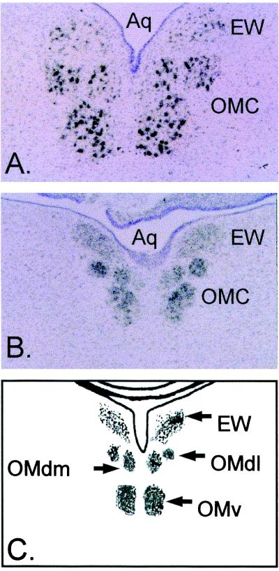
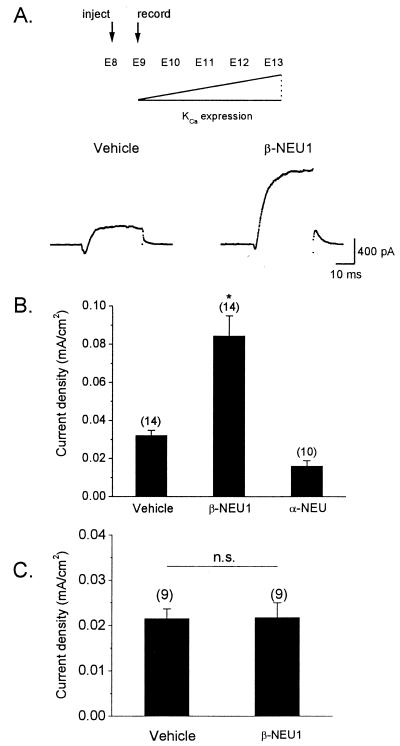
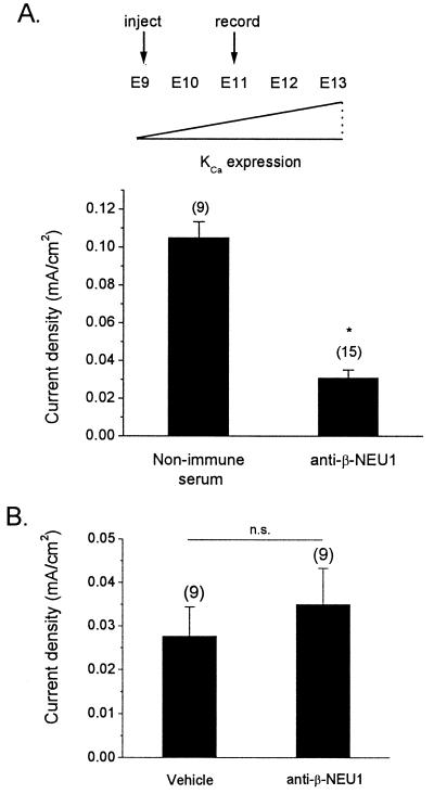
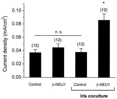
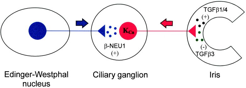
The development of functional Ca(2+)-activated K(+) channels (K(Ca)) in chick ciliary ganglion (CG) neurons requires interactions with afferent preganglionic nerve terminals. Here we show that the essential preganglionic differentiation factor is an isoform of beta-neuregulin-1. beta-Neuregulin-1 transcripts are expressed in the midbrain preganglionic Edinger-Westphal nucleus at developmental stages that coincide with or precede the normal onset of macroscopic K(Ca) in CG neurons. Injection of beta-neuregulin-1 peptide into the brains of developing embryos evoked a robust stimulation of functional K(Ca) channels at stages before the normal appearance of these channels in CG neurons developing in vivo. Conversely, injection of a neutralizing antiserum specific for beta-neuregulin-1 inhibited the development of K(Ca) channels in CG neurons. Low concentrations of beta-neuregulin-1 evoked a robust increase in whole-cell K(Ca) in CG neurons cocultured with iris target tissues. By contrast, culturing CG neurons with iris cells or low concentrations of beta-neuregulin-1 by themselves was insufficient to stimulate K(Ca). These data suggest that the preganglionic factor required for the development of K(Ca) in ciliary ganglion neurons is an isoform of beta-neuregulin-1, and that this factor acts in concert with target-derived trophic molecules to regulate the differentiation of excitability.


Figures

Figure 1
Figure 1
Expression of β-Neu1 transcripts in developing chick oculomotor nuclei. Bright-field micrographs of in situ hybridization histochemistry with antisense riboprobes directed against EGF-domains conserved in all β-NEU1 isoforms. Sections were counterstained with cresyl violet. Note robust hybridization signal in the Edinger–Westphal nucleus (EW) and oculomotor complex (OMC) of E13 (A) and E9 (B) chick embryos. (C) The relative locations of midbrain oculomotor nuclei. The dorsal lateral oculomotor nucleus (OMdl), dorsal medial oculomotor nucleus (OMdm), ventral oculomotor nucleus (OMv), and mesencephalic aqueduct (Aq).
Figure 2
Figure 2
Stimulation of functional KCa by β-NEU1 in CG neurons developing in vivo. (A) Recombinant β-NEU1 peptide, recombinant α-neuregulin peptide, or saline vehicle was injected into the brains of chick embryos at E8. KCa was determined by whole-cell recordings from CG neurons isolated acutely at E9, at stage at which functional KCa is normally present at low levels (Upper). Representative whole-cell KCa currents in E9 CG neurons dissociated from vehicle-injected (Lower Left) and β-NEU1-injected (Lower Right) embryos are shown below. Traces shown are net Ca2+-dependent K+ currents obtained by digital subtraction of currents evoked in normal and Ca2+-free bath salines. (B) Summary of data obtained from several experiments. Injection of β-NEU1 evoked a robust and significant increase in mean KCa density compared to CG neurons isolated from embryos injected with either α-neuregulin or saline vehicle. In this and subsequent figures, bar graphs denote mean ± SEM, numbers above bars denote number of cells tested, and asterisk denotes statistical significance (P < 0.05). (C) Injection of β-NEU1 has no effect on the density or gating of voltage-activated Ca2+ currents.
Figure 3
Figure 3
In vivo injection of β-NEU1-neutralizing antiserum inhibits the normal development of KCa in CG neurons. (A) β-NEU1-neutralizing antiserum, saline vehicle, or nonimmune serum were injected into the brains of E9 chick embryos. Density of ionic currents was determined by whole-cell recordings at E11, a stage at which KCa density is normally increased in CG neurons (Upper). CG neurons from embryos injected with β-NEU1-neutralizing antiserum exhibited significantly reduced mean KCa density compared to neurons from embryos injected with nonimmune serum (Lower). (B) In contrast, injection of neutralizing antiserum had no effect on voltage-activated Ca2+ currents in CG neurons.
Figure 4
Figure 4
β-NEU1 permits KCa stimulation by target tissue myotubes in CG neurons developing in vitro. E9 CG neurons were cocultured with iris myotubes for 12 h in the presence or absence of 1 nM β-NEU1 peptide. Additional control neurons were grown in the absence of iris myotubes but in the presence or absence of 1 nM β-NEU1. Robust macroscopic KCa was observed only in CC neurons grown in the presence of iris myotubes and β-NEU1. Iris myotubes or 1 nM β-NEU1 by themselves were insufficient to promote significant macroscopic KCa. CG neurons cultured in the absence of added factors also failed to express significant whole-cell KCa.
Figure 5
Figure 5
Model for the developmental regulation of CG neuron KCa by multiple cell–cell interactions. The data presented in this and previous studies are consistent with a model in which the development of KCa is regulated by the combined actions of at least three differentiation factors: β-NEU1 (stimulatory, derived from preganglionic nerve terminals); TGFβ1/4 (stimulatory, derived from ocular target tissues); and TGFβ3 (inhibitory, also derived from ocular target tissues). The balance of the in vivo activities of these factors is such that disruption of interactions with either the target tissues or the afferent input prevents the normal development of functional plasma membrane KCa.

References

    1. Fischbach G D, Rosen K M. Annu Rev Neurosci. 1997;20:429–458. - PubMed
    1. Gassmann M, Lemke G. Curr Opin Neurobiol. 1997;7:87–92. - PubMed
    1. Falls D L, Rosen K M, Corfas G, Lane W S, Fischbach G D. Cell. 1993;72:801–815. - PubMed
    1. Sandrock A W, Dryer S E, Rosen K M, Gozani S N, Kramer R, Theill L E, Fischbach G D. Science. 1997;276:599–603. - PubMed
    1. Yang X, Kuo Y, Devay P, Yu C, Role L. Neuron. 1998;20:255–270. - PubMed

Publication types

LinkOut - more resources