. 2001 Feb 27;98(5):2871-6.
doi: 10.1073/pnas.041611398.

Inhibition of caspase 1 reduces human myocardial ischemic dysfunction via inhibition of IL-18 and IL-1beta

Affiliations

Inhibition of caspase 1 reduces human myocardial ischemic dysfunction via inhibition of IL-18 and IL-1beta

B J Pomerantz et al. Proc Natl Acad Sci U S A. .

Abstract

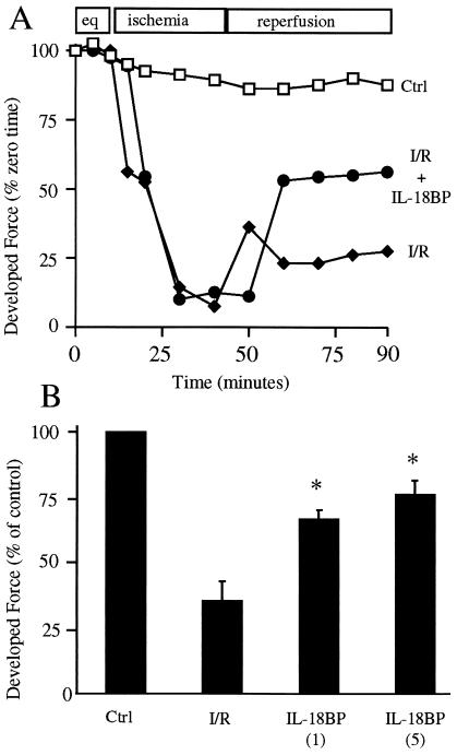
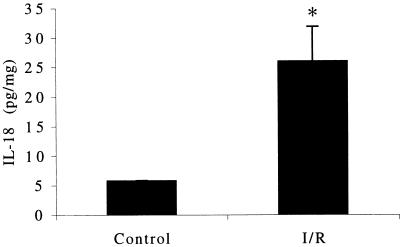
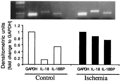
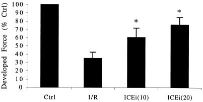
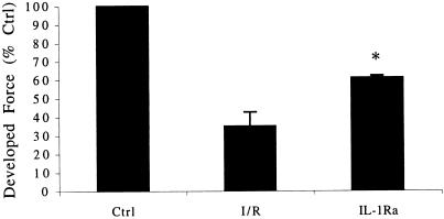
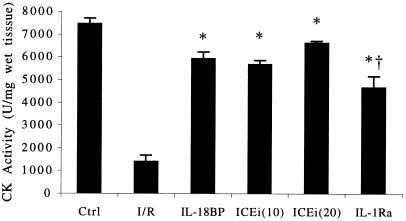
The proinflammatory cytokine IL-18 was investigated for its role in human myocardial function. An ischemia/reperfusion (I/R) model of suprafused human atrial myocardium was used to assess myocardial contractile force. Addition of IL-18 binding protein (IL-18BP), the constitutive inhibitor of IL-18 activity, to the perifusate during and after I/R resulted in improved contractile function after I/R from 35% of control to 76% with IL-18BP. IL-18BP treatment also preserved intracellular tissue creatine kinase levels (by 420%). Steady-state mRNA levels for IL-18 were elevated after I/R, and the concentration of IL-18 in myocardial homogenates was increased (control, 5.8 pg/mg vs. I/R, 26 pg/mg; P < 0.01). Active IL-18 requires cleavage of its precursor form by the IL-1beta-converting enzyme (caspase 1); inhibition of caspase 1 also attenuated the depression in contractile force after I/R (from 35% of control to 75.8% in treated atrial muscle; P < 0.01). Because caspase 1 also cleaves the precursor IL-1beta, IL-1 receptor blockade was accomplished by using the IL-1 receptor antagonist. IL-1 receptor antagonist added to the perifusate also resulted in a reduction of ischemia-induced contractile dysfunction. These studies demonstrate that endogenous IL-18 and IL-1beta play a significant role in I/R-induced human myocardial injury and that inhibition of caspase 1 reduces the processing of endogenous precursors of IL-18 and IL-1beta and thereby prevents ischemia-induced myocardial dysfunction.


Figures

Figure 1
Figure 1
Effect of IL-18BP on ischemia-induced myocardial contractile dysfunction. (A) Kinetic response to ischemic injury. After equilibration (eq), control (Ctrl) trabeculae were suprafused under normoxic conditions throughout the experiment. Trabeculae were subjected to I/R in the absence or presence of IL-18BP (5 μg/ml) as described in the experimental model. The vertical axis indicates percent of developed force compared with initiation of the experiment (time 0). The data are derived from trabeculae of a single patient and are representative of the methods used to calculate the mean change in developed force at 90 min. (B). Postischemic developed force after neutralization of IL-18 with IL-18BP. Results are expressed as the mean percent change in developed force relative to Ctrl after completion of reperfusion (90 min). Numbers in parentheses indicate IL-18BP in μg/ml (n = 6). *, P < 0.01 compared with I/R.
Figure 2
Figure 2
Myocardial IL-18 protein content. Trabeculae were homogenized after 90 min of suprafusion under normoxic conditions (Control) or 45 min after 30 min of ischemia. Trabeculae were matched from the same subjects. IL-18 levels are indicated on the vertical axis in pg/ml (n = 4). *, P < 0.01.
Figure 3
Figure 3
Steady-state IL-18 and IL-18BP mRNA levels in control and ischemic atrial tissue. Tissues obtain after conditions described in Fig. 2 were snap-frozen in liquid nitrogen and kept frozen at −70°C. After homogenization in Tri-Reagent and mRNA isolation, levels of IL-18 and IL-18BP mRNA were determined by reverse transcription-coupled PCR. Data are from one of two subjects evaluated. GAPDH, glyceraldehyde-3-phosphate dehydrogenase.
Figure 4
Figure 4
Location of IL-18 in human myocardium. Immunohistochemical staining of human atrial tissue before insertion of atrial canula. (Left) Section through an atrial blood vessel. IL-18 is present within vascular endothelial cells (EC) and in resident myocardial macrophages (Mφ) (Right) Section through atrial muscle. Myocytes (Myo) are identified and IL-18 is observed in the resident macrophages. In this staining technique, IL-18 appears pink.
Figure 5
Figure 5
Effect of ICE inhibition on postischemic developed force. Results are expressed as the mean percent change in developed force relative to control (Crtl) after I/R. Numbers in parentheses indicate the concentration of ICEi in μg/ml (n = 7). *, P < 0.01 compared with I/R.
Figure 6
Figure 6
Preservation of contractile function after I/R and blockade of IL-1 receptors with IL-1Ra. Results are expressed as the mean percent change in developed force relative to control (Ctrl) after completion of reperfusion. The concentration of IL-1Ra is 20 μg/ml (n = 5). *, P < 0.01 compared with I/R.
Figure 7
Figure 7
Tissue CK activity after I/R. CK is expressed in units of activity per mg (wet weight of tissue). The experimental conditions are indicated under the horizontal axis. Ctrl and I/R (n = 6); IL-18BP at 5 μg/ml (n = 5); ICEi at 10 and 20 μg/ml (n = 5, each group); IL-1Ra at 20 μg/ml (n = 6). *, P < 0.05 compared with I/R; †, P < 0.05 for ICEi (20) compared with IL-1Ra.

References

    1. Bolli R. Circulation. 1990;82:723–738. - PubMed
    1. Herskowitz A, Choi S, Ansari A A, Wesselingh S. Am J Pathol. 1995;146:419–428. - PMC - PubMed
    1. Meldrum D R, Cleveland J C, Jr, Cain B S, Meng X, Harken A H. Ann Thorac Surg. 1998;65:439–443. - PubMed
    1. Gurevitch J, Frolkis I, Yuhas Y, Paz Y, Matsa M, Mohr R, Yakirevich V. J Am Coll Cardiol. 1996;28:247–252. - PubMed
    1. Cleveland J C J, Meldrum D R, Cain B S, Banerjee A, Harken A H. Circulation. 1997;96:29–32. - PubMed

Publication types

MeSH terms

LinkOut - more resources