Clinical Trial
. 2001 Feb;131(2):167-75.
doi: 10.1016/s0002-9394(00)00732-7.

The age-related eye disease study (AREDS) system for classifying cataracts from photographs: AREDS report no. 4

Clinical Trial

The age-related eye disease study (AREDS) system for classifying cataracts from photographs: AREDS report no. 4

Age-Related Eye Disease Study Research Group. Am J Ophthalmol. 2001 Feb.

Abstract

Purpose: To describe the system for grading cataracts from photographs in the Age-Related Eye Disease Study (AREDS).

Methods: The system for grading cataracts in AREDS uses photographs taken in a standardized fashion with specially modified cameras at 11 clinical centers. The photographs are evaluated by graders for quality and cataract severity at a central reading center. The area of lens involvement is used to assess the severity of cortical and posterior subcapsular opacities. Optical density of nuclear opacity is graded against a series of seven standard photographs. Contemporaneous variability in grading is evaluated periodically by having a second examiner regrade a subset of the photographs. Temporal variability is assessed by annually regrading a subset of photographs.

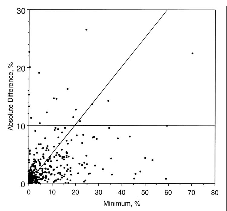
Results: Photographs of 925 eyes, most with no or early lens opacities, were regraded to assess intergrader reliability. For cortical opacities, there was an absolute difference of 10% or greater of area involved in 1.9% of the replicate gradings. For posterior subcapsular opacities an absolute difference of 5% of area involved was noted in 2.8% of the regraded photographs. For nuclear opacities, absolute differences of 1.5 or more steps were observed in 0.6% of eyes. There was little evidence of temporal drift in grading any of the three types of opacity during four annual regrades.

Conclusions: We have demonstrated a high degree of reliability in grading the severity of lens opacities in a large study cohort with mostly early lens changes, the type of cohort most likely to be entered in clinical trials involving cataract prevention. The Age-Related Eye Disease Study System for Classifying Cataracts From Photographs could be useful in studies where there is a need to standardize data collection over time and across different data collection sites. Limitations of the system include the cost of implementation and, currently, the limited amount of data on grading reproducibility for more advanced lens opacities.


Figures

FIGURE 1.
FIGURE 1.
Standard photographs 1 (no opacity) through 7 (extremely severe opacity) for grading nuclear opacities. A-G represent standard photographs 1-7.
FIGURE 2.
FIGURE 2.
Grid used with retroillumination photographs to grade cortical and posterior subcapsular opacities.
FIGURE 3.
FIGURE 3.
(Left) Retroillumination photograph of a cortical opacity. (Right) Retroillumination photograph of cortical opacity with overlying grid. The cortical opacity occupies 12% of the area within the central two circles of the grid (central 5 mm of the lens) and 25% of the full visible lens.
FIGURE 4.
FIGURE 4.
(Left) Retroillumination photograph of a posterior subcapsular opacity. (Right) Retroillumination photograph of a posterior subcapsular opacity with overlying grid. The posterior subcapsular opacity occupies 15% of the area within the central two circles of the grid (central 5 mm of the lens).
FIGURE 5.
FIGURE 5.
Comparison of two contemporaneous grades of cortical opacity from retroillumination photographs (n = 925 eyes). Scatter plot of minimum grade compared with the absolute difference in grades. Cortical opacity grade is the percent of involvement by the opacity of the central 5-mm diameter circle of the lens. The points above the horizontal reference line have an absolute difference in grades of more than 10%. Points above the diagonal line have a relative difference of more than 50%. Intraclass correlation coefficient = 0.94.
FIGURE 6.
FIGURE 6.
Comparison of two contemporaneous grades of posterior subcapsular opacity from retroillumination photographs (n = 925 eyes). Scatter plot of minimum grade compared with the absolute difference in grades. The posterior subcapsular opacity grade is the percent of involvement by the opacity of the central 5-mm diameter circle of the lens. Points above the horizontal reference line have an absolute difference in grades of more than 5%. Intraclass correlation coefficient = 0.82.
FIGURE 7.
FIGURE 7.
Comparison of two contemporaneous grades of nuclear opacity from slit-lamp photographs (n = 925 eyes). Scatter plot of minimum grade compared with the absolute difference in grades. Nuclear opacity grade ranges from 1.0 to 5.0, with respect to standard photographs of nuclear opacity. Points above the horizontal reference line have an absolute difference in grades of more than 1.5 units. Intraclass correlation coefficient = 0.90.
FIGURE 8.
FIGURE 8.
Comparison of baseline grades with each of four annual regrades. Box plots of baseline grade minus regrade. Positive differences indicate regrades lower than baseline. In a box plot, the horizontal line in the box is the median; the lower (upper) edge of the box is the 25th (75th) percentile; the lower and upper vertical lines extend to the fifth and 95th percentiles; the distinct points indicate extreme differences that are in the lower or upper 5% of values. The off-center location of the median in the box or vertical lines of unequal length indicate an asymmetrical distribution. A cortical opacity, B posterior subcapsular opacity, C nuclear opacity.

References

    1. Klein BEK, Klein R, Linton KLP, Magli YL, Neider M. Assessment of cataracts from photographs in the Beaver Dam Eye Study. Ophthalmology. 1990;97:1428–1433. - PubMed
    1. Magli YL, Klein BEK, Sperduto RD, Hubbard LD, Neider MW, King WN, Davis MD, Age-Related Eye Disease Study (AREDS) Research Group AREDS extension of the Wisconsin Lens Opacity Grading System. Invest Ophthalmol Vis Sci. 1997;38:S177.
    1. Age-Related Eye Disease Study Research Group The Age-Related Eye Disease Study (AREDS): Design implications. AREDS Report No. 1. Controlled Clin Trials. 1999;20:573–600. - PMC - PubMed

Publication types