Comparative Study
. 2001:1:3.
doi: 10.1186/1471-213x-1-3. Epub 2001 Feb 14.

The mouse anterior chamber angle and trabecular meshwork develop without cell death

Affiliations
Comparative Study

The mouse anterior chamber angle and trabecular meshwork develop without cell death

R S Smith et al. BMC Dev Biol. 2001.

Abstract

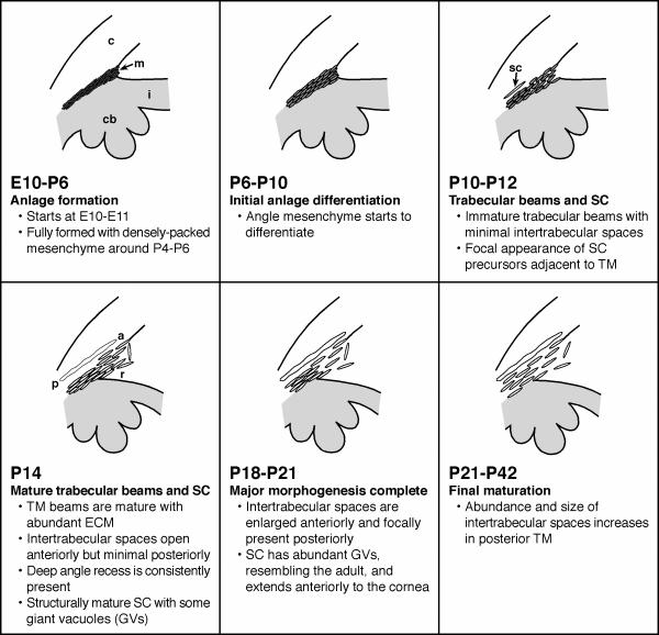
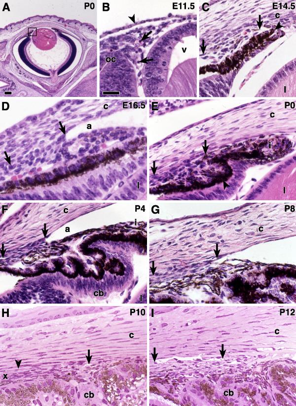
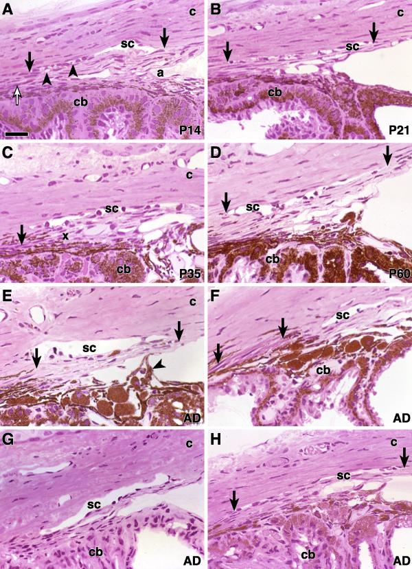
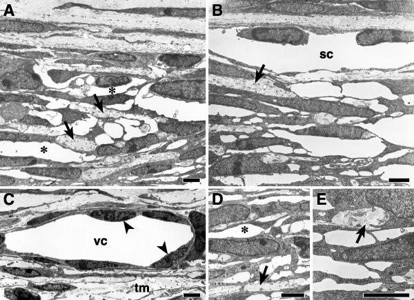
Background: The iridocorneal angle forms in the mammalian eye from undifferentiated mesenchyme between the root of the iris and cornea. A major component is the trabecular meshwork, consisting of extracellular matrix organized into a network of beams, covered in trabecular endothelial cells. Between the beams, channels lead to Schlemm's canal for the drainage of aqueous humor from the eye into the blood stream. Abnormal development of the iridocorneal angle that interferes with ocular fluid drainage can lead to glaucoma in humans. Little is known about the precise mechanisms underlying angle development. There are two main hypotheses. The first proposes that morphogenesis involves mainly cell differentiation, matrix deposition and assembly of the originally continuous mesenchymal mass into beams, channels and Schlemm's canal. The second, based primarily on rat studies, proposes that cell death and macrophages play an important role in forming channels and beams. Mice provide a potentially useful model to understand the origin and development of angle structures and how defective development leads to glaucoma. Few studies have assessed the normal structure and development of the mouse angle. We used light and electron microscopy and a cell death assay to define the sequence of events underlying formation of the angle structures in mice.

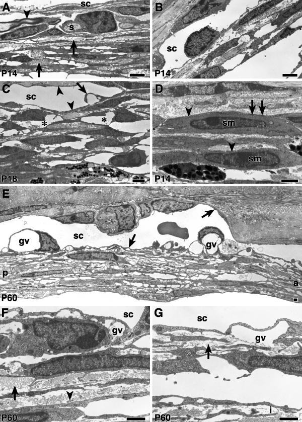
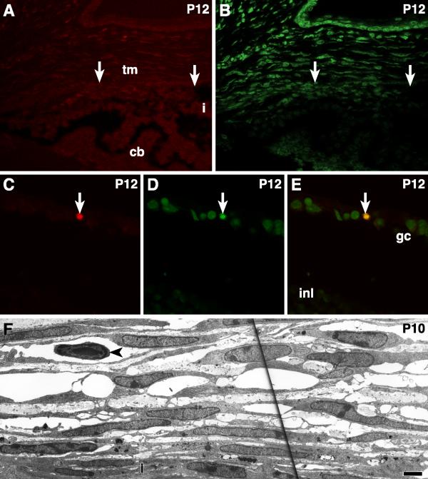
Results: The mouse angle structures and developmental sequence are similar to those in humans. Cell death was not detectable during the period of trabecular channel and beam formation.

Conclusions: These results support morphogenic mechanisms involving organization of cellular and extracellular matrix components without cell death or atrophy.


Figures

Figure 1
Figure 1
Formation of the mouse iridocorneal angle. A diagrammatic representation of iridocorneal angle morphogenesis is shown. c = cornea, cb = ciliary body, i = iris, m = angle mesenchyme, sc = Schlemm's canal, r = deep angle recess. a = anterior, p = posterior.
Figure 2
Figure 2
Iridocorneal angle E11.5 to P12 Images from paraffin (A -G) and plastic (G-H) embedded B6 eyes of the indicated ages. (A) The box indicates the iridocorneal angle region that is illustrated at high power in the other panels of Figures 1 and 2. (B) At E11.5, loose mesenchymal tissue is present between the anterior edge of the optic cup (oc), the lens vesicle (v), and the surface ectoderm (arrowhead). Primitive vascular channels contain nucleated red blood cells (arrows). (C) At E14.5, two layers of epithelium form the OC region that will develop into the iris and ciliary body. The anterior layer is heavily pigmented (arrowhead). The arrows indicate the anterior and posterior extent of undifferentiated angle mesenchyme. The cornea (c) and lens (l) are well defined. (D) At E16.5, a small angle recess is present (a). The location of the future TM is evident (arrows). (E) In a newborn mouse, the mesenchyme of the developing iris (i) and TM (arrows) regions are distinguishable. The TM cells have elongated, more densely-staining nuclei and are arranged in lamellae (arrows). The ciliary processes (arrowheads) have begun to form. The angle recess is artifactually compressed in this image. (F) At P4, there is a long angle recess (a), and the iris and ciliary body (cb) are well formed. The cells of the future TM (arrows) show a dense lamellar arrangement. (G) At P8, the developing TM is less compressed than at earlier ages (arrows). (H) At P10, an endothelial lined vascular channel (arrowhead) is present at some locations. Intertrabecular spaces have begun to open in the anterior portion of the TM (arrow). The posterior aspect of the TM remains compressed (x). (I) At P12, A well-formed SC (arrows) is easily identified exterior to the posterior TM. Internal to SC, both anterior and posterior meshwork has become more open. Bars 200 μm (A) and 40 μm (B-I).
Figure 3
Figure 3
Iridocorneal angle P14 to P63 Hematoxylin and eosin stained plastic sections from mice of the indicated ages. (A-D) strain B6. ( A) At P14, SC (arrows) contains vacuolar structures (arrowheads) that were confirmed to be giant vacuoles by EM (see below). The developing ciliary muscle is characterized by eosinophilic cytoplasm (open arrow). Intertrabecular spaces are obvious in the anterior TM and the deep angle recess (a) is present as a space between the anterior TM and iris root. c = cornea, cb = ciliary body. ( B) By P21, SC (arrows) extends from the posterior ciliary body to the end of Descemet's membrane. There are large spaces in the anterior TM. (C) By P35 there is further opening of the intertrabecular spaces that extend more posteriorly. The posterior TM (x) remains closely attached to the ciliary body, as it does in the adult. The ciliary muscle (arrow) consists of a few muscle fibers. (D) This P60 eye has a well developed SC (arrows) and TM and is very similar to that shown for P35. Comparison to older mice (up to 1 year old, not shown) indicates that the iridocorneal angle has reached maturity. The adult structure is similar in other mouse strains (E-H). All of the adult mice were approximately 63 days old. (E) A 129/SvEvTac mouse has a robust TM (arrows) and a broad SC. An iris process attaches to the anterior TM (arrowhead). (F) In this 129BS mouse, there is a robust TM and SC. The ciliary muscle (arrows) is particularly prominent in this strain. (G) BALB/cByJ. (H) In this DBA/2J mouse, SC is present but shows mild artifactual compression (arrows). Bar 40 μm.
Figure 4
Figure 4
Ultrastructure of the TM and SC in B6 mice at P10 (A, D) In the posterior TM, spaces are developing between the trabecular beams (asterisks). Small amounts of collagen and elastic tissue are demonstrated within the beams (arrows). Schlemm's canal is absent in these sections. (B, E) In the anterior TM, the spaces between adjacent trabecular beams are generally larger than posteriorly (compare B and E that are anterior to A and D that are posterior). SC is present in B but giant vacuoles are absent. Elastic tissue and collagen (arrows) are present in amounts similar to that of the posterior meshwork. (C) In a different level of section to B, SC is represented by a vascular channel (vc) adjacent to the differentiating TM (tm). The endothelial cells (arrowheads) lining this channel are less attenuated than in the adult SC and giant vacuoles are absent. Bars 1 μm.
Figure 5
Figure 5
Ultrastructure of the TM and SC in B6 mice from P14 to P60 ( A, B, D) are from the same P14 mouse. (A) At P14 the spaces in the posterior TM are smaller than at older ages (compare to a P18 eye in C). Trabecular beam collagen and elastic tissue (arrows) is more abundant than at younger ages. A Schwann cell (s) and accompanying myelinated nerve (arrowhead) are present close to SC. (B) In the anterior TM, there is a well-developed SC, but giant vacuoles are not common. There are fewer trabecular beams and larger intertrabecular spaces than in the posterior TM (compare to A, and see F, G). (C) The spaces (asterisks) between trabecular beams in this region of the posterior TM are more extensive than at P14. SC is lined by a thin endothelium (arrowheads) and contains giant vacuoles (arrow). (D) Smooth muscle cells (sm) lie internal to Schlemm's canal near its posterior termination. They are characterized by pinocytotic vesicles near the cell membrane (arrowheads), focal density of the plasma membrane (arrows) and cytoplasmic filaments (not seen at this magnification). (E) At P60, SC is lined by attenuated endothelium (arrows) and contains giant vacuoles (gv). a = anterior, p = posterior. (F) Partial segment of the posterior TM at P60. The trabecular beam extracellular matrix is dense. Collagen (arrow) is abundant while elastic tissue (arrowhead) is relatively sparse. (G) In the anterior TM, the beams are more delicate, and contain less extracellular matrix (arrow). A portion of the anterior iris (i) is present in this image. Bars 1 μm.
Figure 6
Figure 6
Absence of cell death in the developing iridocorneal angle. A double labeling assay that identifies fragmented DNA using fluorescently labeled dUTP (A, C) and detects chromatin condensation by binding of the dye YOYO-1 (B, D) was used to detect programmed cell death (PCD). Both assays were negative in a P12, B6 iridocorneal angle (A, B). The same was true for many sections at ages that spanned angle morphogenesis. i= iris, cb = ciliary body, arrows indicate the extremities of the TM (tm). (C, D, E) A cell undergoing PCD (arrow) is identified by double labeling in the retinal ganglion cell layer (gc) of the same eye shown in A and B. inl = inner nuclear layer. Dying retinal ganglion cells (RGCs) acted as internal positive controls for the PCD assays. Testis sections served as additional positive controls with each batch of processed slides, and abundant apoptotic cells were always detected. (F) Morphologic features of cell death were absent in the TM of a P10, B6 mouse. The trabecular cells demonstrate normal nuclei and normal cytoplasmic morphology. The same was true in many sections of eyes of different ages and strains. The iris (i) is resting against the inner edge of this central portion of the TM. A small lymphocyte (arrowhead) lies in the space between two trabecular beams. Bar 1 μm.

References

    1. Ritch R, Shields MB, Krupin T. The Glaucomas, Clinical Science, 2nd edn. St Louis, MO: Mosby-Year Book; 1996.
    1. Caprioli J. The ciliary epithelia and aqueous humor. Adler's Physiology of the Eye Edited by William M. Hart J, 9th ed. pp. 228-247. St. Louis: Mosby Year Book; 1992. pp. 228–247.
    1. Hart WM. Intraocular Pressure. Adler's Physiology of the Eye Edited by William M. Hart J, 9th ed. pp. 248-267. St. Louis: Mosby Year Book; 1992. pp. 248–267.
    1. Bill A. Uveoscleral drainage of aqueous humor: physiology and pharmacology. Prog Clin Biol Res. 1989;312:417–427. - PubMed
    1. Mann IC. The Development of the Human Eye, First edn. Cambridge: Cambridge University Press; 1928.

Publication types

MeSH terms

LinkOut - more resources