. 2001 Mar 1;20(5):998-1009.
doi: 10.1093/emboj/20.5.998.

Recruitment, activation and retention of caspases-9 and -3 by Apaf-1 apoptosome and associated XIAP complexes

Affiliations

Recruitment, activation and retention of caspases-9 and -3 by Apaf-1 apoptosome and associated XIAP complexes

S B Bratton et al. EMBO J. .

Abstract

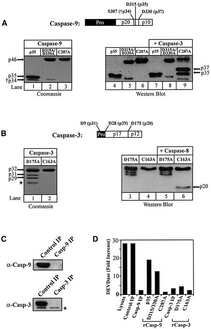
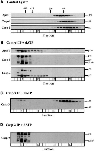
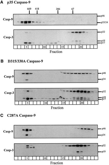
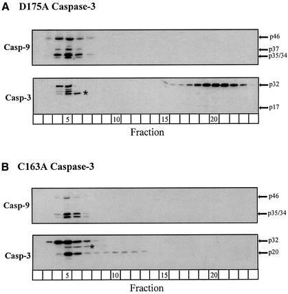
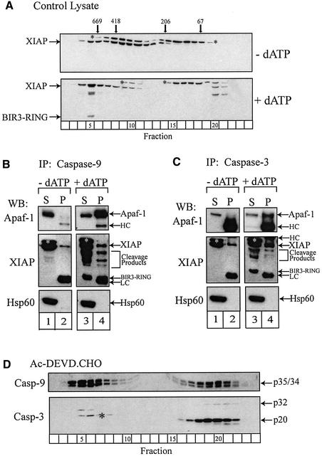
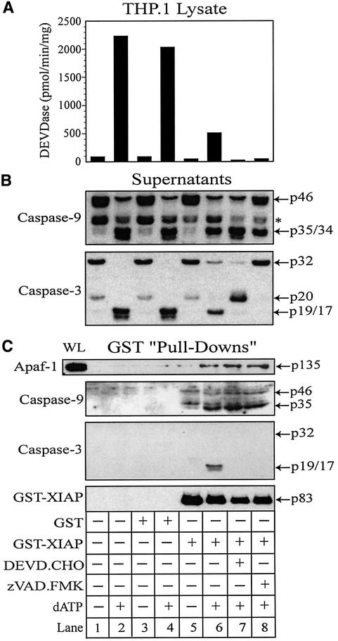
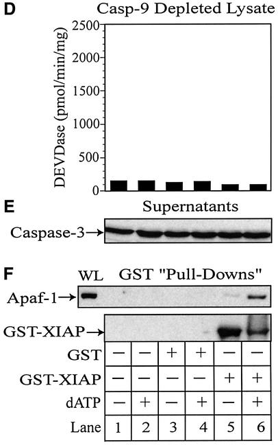
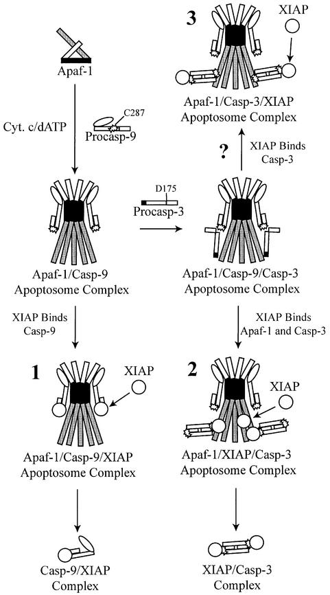
During apoptosis, release of cytochrome c initiates dATP-dependent oligomerization of Apaf-1 and formation of the apoptosome. In a cell-free system, we have addressed the order in which apical and effector caspases, caspases-9 and -3, respectively, are recruited to, activated and retained within the apoptosome. We propose a multi-step process, whereby catalytically active processed or unprocessed caspase-9 initially binds the Apaf-1 apoptosome in cytochrome c/dATP-activated lysates and consequently recruits caspase-3 via an interaction between the active site cysteine (C287) in caspase-9 and a critical aspartate (D175) in caspase-3. We demonstrate that XIAP, an inhibitor-of-apoptosis protein, is normally present in high molecular weight complexes in unactivated cell lysates, but directly interacts with the apoptosome in cytochrome c/dATP-activated lysates. XIAP associates with oligomerized Apaf-1 and/or processed caspase-9 and influences the activation of caspase-3, but also binds activated caspase-3 produced within the apoptosome and sequesters it within the complex. Thus, XIAP may regulate cell death by inhibiting the activation of caspase-3 within the apoptosome and by preventing release of active caspase-3 from the complex.


Figures

None
Fig. 1. Reconstitution of immunodepleted lysates with recombinant wild-type and mutant caspases-9 and -3. (A) Recombinant wild-type and fully processed caspase-9 (lane 1), non-cleavable unprocessed D315/330A caspase-9 (lane 2) and catalytically inactive C287A caspase-9 (lane 3) were isolated and purified from bacteria and their purity was determined by Coomassie Blue staining. Each caspase-9 protein was incubated with (lanes 7–9) or without (lanes 4–6) recombinant active caspase-3 (100 nM) for 1 h at 37°C. The samples were subsequently separated by SDS–PAGE and immunoblotted using a polyclonal anti-caspase-9 antibody. (B) The non-cleavable D175A caspase-3 (lanes 1, 3 and 5) and catalytically inactive C163A caspase-3 (lanes 2, 4 and 6) mutants were purified from bacteria, Coomassie-stained to assess purity and exposed to purified recombinant caspase-8 (100 nM) for 1 h at 37°C. In the D175A caspase-3 preparation, two minor internal translation products of ∼27 and ∼29 kDa were present (Fernandes-Alnemri et al., 1996), as well as a minor bacterial contaminant (*). (C) THP.1 lysates were immunodepleted of caspase-9 or -3 and (D) reconstituted with their corresponding wild-type or mutant caspases (200 nM). The reconstituted lysates were dATP-activated for 1 h at 37°C and assayed for DEVDase activity as described in Materials and methods. The anti-caspase-3 antibody recognized a non-specific band (*) in lysates, which served as a fortuitous loading control.
None
Fig. 2. The Apaf-1 apoptosome sequentially recruits, activates and retains caspases-9 and -3. (A) Unactivated control lysates, (B) dATP-activated immunoprecipitated (IP) control lysates, (C) dATP-activated caspase-9-depleted lysates or (D) dATP-activated caspase-3-depleted lysates were fractionated by gel filtration, as described in Materials and methods. Each fraction was mixed with 10× SDS loading buffer, separated by SDS–PAGE and immunoblotted for Apaf-1, caspase-9 and/or caspase-3. The arrows represent either the unprocessed or processed form(s) of the caspases; a non-specific band (*) was detected by the anti-caspase-3 antibody.
None
Fig. 3. Recruitment of caspase-3 to the Apaf-1 apoptosome requires catalytically active processed or unprocessed caspase-9. Caspase-9-depleted lysates were reconstituted with (A) fully processed wild-type p35 caspase-9, (B) non-cleavable D315/330A caspase-9 or (C) catalytically inactive C287A caspase-9. Following dATP activation for 1 h at 37°C, the reconstituted lysates were fractionated and immunoblotted for caspases-9 and -3, as described in Materials and methods. The arrows represent either the unprocessed or processed form(s) of the caspases; a non-specific band (*) was detected by the anti-caspase-3 antibody.
None
Fig. 4. Caspase-9 recruits caspase-3 to the Apaf-1 apoptosome via recognition of a critical aspartate (D175) residue. Caspase-3-depleted lysates were reconstituted with (A) unprocessable D175A caspase-3 or (B) catalytically inactive C163A caspase-3. Following dATP activation for 1 h at 37°C, the reconstituted lysates were fractionated and immunoblotted for caspases-9 and -3, as described in Materials and methods. The arrows represent either the unprocessed or processed form(s) of the caspases; a non-specific band (*) was detected by the anti-caspase-3 antibody.
None
Fig. 5. Co-immunoprecipitation of Apaf-1, caspase-9, caspase-3 and XIAP. (A) Control and dATP-activated THP.1 lysates were fractionated by gel filtration. The fractions were separated by SDS–PAGE and immunoblotted for XIAP using an antibody raised against its C-terminus. Control and dATP-activated lysates (∼15 mg/ml) were immunoprecipitated with an antibody against (B) caspase-9 or (C) caspase-3 and the resulting immuno complexes were recovered by centrifugation. The supernatants (S) and washed immunocomplexes (P) were separated by SDS–PAGE and western blotted (WB) for Apaf-1, XIAP or Hsp60. (D) Lysates were pretreated with DEVD⋅CHO (200 nM) for 1 h at 4°C and subsequently dATP-activated at 37°C for 1 h. The lysates were fractionated by gel filtration and analyzed by SDS–PAGE/immunoblotting for caspases-9 and -3, as described in Materials and methods. The arrows represent either the full-length proteins or, in the case of XIAP, various cleavage products. Bands corresponding to the light chain (LC) or heavy chain (HC) of the immunoprecipitated antibodies are also shown. Separation of the LC and the BIR3-RING fragment was difficult, but this fragment was only apparent in caspase-9 immunocomplexes obtained from dATP-activated lysates. Non-specific bands (*) were detected by the XIAP or caspase-3 antibodies.
None
Fig. 6. XIAP directly associates with Apaf-1 and retains active caspase-3 within the apoptosome. THP.1 lysates were activated with dATP (lanes 2, 4, 6, 7 and 8) for 1 h at 37°C in the presence of GST (lanes 3 and 4) or GST–XIAP (lanes 5–8). In some treatments, the lysate was pretreated for 15 min with DEVD⋅CHO (200 nM) (lane 7) or z-VAD⋅FMK (25 µM) (lane 8). A small aliquot of each sample was immediately assayed for (A) DEVDase activity and the remainder incubated with GSH–Sepharose beads (20 µl) overnight at 4°C. Following centrifugation, supernatants were collected and the beads thoroughly washed (four times) with assay buffer. (B) Supernatants and (C) GST–XIAP/protein complexes were subsequently mixed with 2× SDS loading buffer, separated by SDS–PAGE and immunoblotted for Apaf-1, caspase-9, caspase-3 and/or XIAP. Similarly, caspase-9-depleted lysates were activated with dATP (lanes 2, 4 and 6) in the presence of GST (lanes 3 and 4) or GST–XIAP (lanes 5 and 6). A small aliquot of each sample was assayed for (D) DEVDase activity and the remainder incubated with GSH–Sepharose beads (20 µl) overnight at 4°C. After centrifugation, the resulting (E) supernatants and (F) GST–XIAP/protein complexes were separated by SDS–PAGE and immunoblotted for Apaf-1, caspase-3 and/or XIAP, as described above. All Apaf-1 western blots are presented at the same exposures; ‘WL’ represents the total amount of Apaf-1 present in each whole lysate that was available to interact with GST–XIAP.
None
Fig. 7. Models for sequential recruitment and activation of caspases-9 and -3 and retention by XIAP. Apaf-1 undergoes cytochrome c/dATP-dependent oligomerization and recruits caspase-9 to the apoptosome through CARD–CARD interactions. Active (pro)caspase-9 then associates with XIAP (1) and/or recruits caspase-3 to the apoptosome through interactions involving the C287 residue in caspase-9 and the D175 residue in caspase-3. Following its activation, active caspase-3 is retained within the apoptosome via an interaction with XIAP, which is simultaneously associated with Apaf-1 (2).

References

    1. Bratton S.B., MacFarlane,M., Cain,K. and Cohen,G.M. (2000) Protein complexes activate distinct caspase cascades in death receptor and stress-induced apoptosis. Exp. Cell Res., 256, 27–33. - PubMed
    1. Cain K., Brown,D.G., Langlais,C. and Cohen,G.M. (1999) Caspase activation involves the formation of the aposome, a large (∼700 kDa) caspase-activating complex. J. Biol. Chem., 274, 22686–22692. - PubMed
    1. Cain K., Bratton,S.B., Langlais,C., Walker,G., Brown,D.G., Sun,X.M. and Cohen,G.M. (2000) Apaf-1 oligomerizes into biologically active ∼700 kDa and inactive ∼1.4 MDa apoptosome complexes. J. Biol. Chem., 275, 6067–6070. - PubMed
    1. Clem R.J. and Miller,L.K. (1994) Control of programmed cell death by the baculovirus genes p35 and iap. Mol. Cell. Biol., 14, 5212–5222. - PMC - PubMed
    1. Cohen G.M. (1997) Caspases: the executioners of apoptosis. Biochem. J., 326, 1–16. - PMC - PubMed

Publication types