. 2001 Mar 1;20(5):1010-9.
doi: 10.1093/emboj/20.5.1010.

Arabidopsis glucosidase I mutants reveal a critical role of N-glycan trimming in seed development

Affiliations

Arabidopsis glucosidase I mutants reveal a critical role of N-glycan trimming in seed development

M Boisson et al. EMBO J. .

Abstract

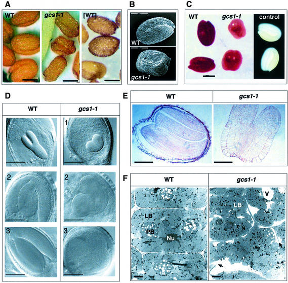
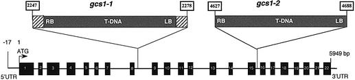
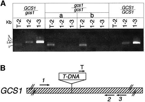
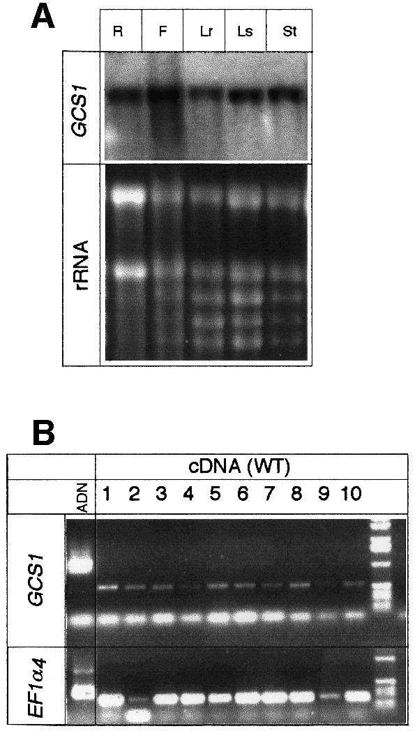
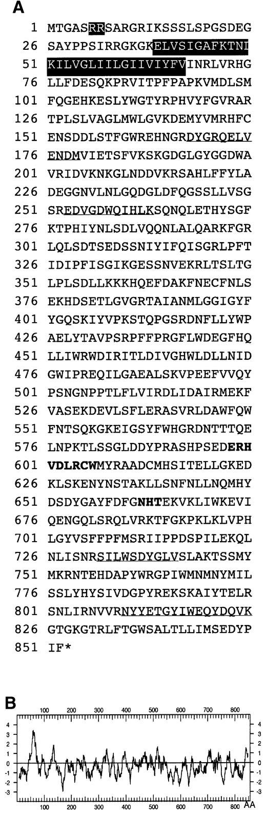
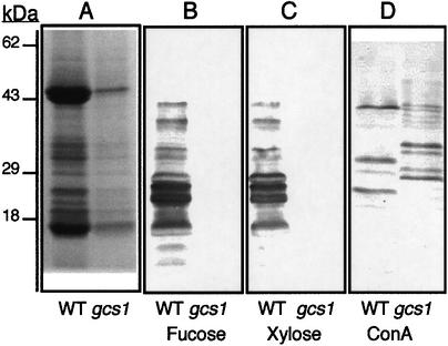
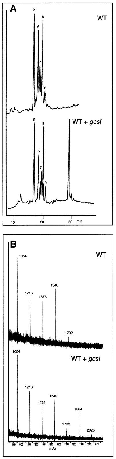
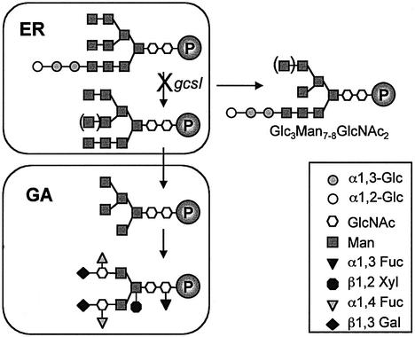
Glycoproteins with asparagine-linked (N-linked) glycans occur in all eukaryotic cells. The function of their glycan moieties is one of the central problems in contemporary cell biology. N-glycosylation may modify physicochemical and biological protein properties such as conformation, degradation, intracellular sorting or secretion. We have isolated and characterized two allelic Arabidopsis mutants, gcs1-1 and gcs1-2, which produce abnormal shrunken seeds, blocked at the heart stage of development. The mutant seeds accumulate a low level of storage proteins, have no typical protein bodies, display abnormal cell enlargement and show occasional cell wall disruptions. The mutated gene has been cloned by T-DNA tagging. It codes for a protein homologous to animal and yeast alpha-glucosidase I, an enzyme that controls the first committed step for N-glycan trimming. Biochemical analyses have confirmed that trimming of the alpha1,2- linked glucosyl residue constitutive of the N-glycan precursor is blocked in this mutant. These results demonstrate the importance of N-glycan trimming for the accumulation of seed storage proteins, the formation of protein bodies, cell differentiation and embryo development.


Figures

None
Fig. 1. Microscopy analyses of wild-type and mutant seeds. (A) Light microscopy analyses of wild-type seeds (WT), gcs1 seeds selected from a heterozygous plant (gcs1-1) and seeds produced by a plant homozygous for the gcs1 mutation, which ectopically expresses GCS1 cDNA ([WT]). Scale bars, 200 µm. (B) Electron microscopy pictures of wild-type (top) and gcs1 mutant seeds (bottom). Scale bars, 200 µm. (C) Embryo viability using the tetrazolium test. Results shown are obtained with wild-type embryo (WT), gcs1-1 embryo (gcs1-1) and wild-type embryo boiled for 30 min as a negative control. Since the negative control is completely white, it is shown on a dark background. Scale bar, 200 µm. (D) Cleared seed viewed with Nomarski optics. Comparison of the development of the wild-type (WT) and gcs1 embryos after the heart stage. The radicle and cotyledons of gcs1-1 do not elongate to form the torpedo embryo (1). However, gcs1-1 embryos enlarge and fill the whole mature seed (2 and 3). Scale bars, 100 µm. (E) Light microscopy analyses of semi-thin sections stained with toluidin blue. Mature wild-type (WT) and gcs1-1 seeds. Scale bars, 80 µm. (F) Transmission electron micrographs of representative cotyledon cells from the wild-type and mutant embryos. LB, lipid bodies; Nu, nucleus; PB, protein bodies; V, vacuoles. Arrows indicate incomplete cell walls occasionally observed in the mutant cells. Scale bars, 1 µm.
None
Fig. 2. Molecular analyses. Schematic representation of the gene structure at the GCS1 locus, in wild-type and gcs1 mutant genomes. The structure of the gene is deduced from the comparison between genomic and cDNA sequences (DDBJ/EMBL/GenBank accession No. AJ278990). In gcs1-1, the T-DNA was inserted between nucleotides 2247 and 2278, leading to a small deletion of 30 bp. Dashed boxes represent insertions of 26 and 22 bp of unknown origin in gcs1-1. In gcs1-2, the T-DNA was inserted between nucleotides 4629 and 4698, leading to a deletion of 58 bp. Black boxes are exons. The 5′UTR was obtained by 5′-RACE–PCR.
None
Fig. 3. (A) PCR analyses of the structure of the GCS1 locus. The DNA fragments corresponding to GCS1 loci were PCR amplified with genomic DNA extracted from individual embryos, after removing the seed coat. The results shown have been obtained with DNA from embryos produced by heterozygous (GCS1/gcs1) or wild-type (GCS1/GCS1) plants and with two representative selected mutant embryos (gcs1/gcs1, a and b). (B) Schematic representation of the insertion locus and location of the primers used for the analyses.
None
Fig. 4. Gene expression analyses. (A) Northern blot analysis of GCS1 gene expression in different tissues: roots (R), flower (F), rosette leaves (Lr), stem leaves (Ls) and stem (St). Eight micrograms of total RNA were loaded per lane. (B) RT–PCR analyses of GCS1 gene expression during silique development. Total mRNAs were extracted at different stages of flower (lane 1) and silique development (lanes 2–10). Siliques were taken in pairs from the top of the main axis, starting with the first siliques emerging from the flower (lane 2), to dry mature siliques (lane 10). The EF-1α gene (Liboz et al., 1990) was included as a control. To control that no genomic DNA contaminated PCR products, oligonucleotides were designed to amplify a region overlapping an intron. The genomic control is presented for both GCS1 and EF-1α genes.
None
Fig. 5. Structure of the GCS1 protein. (A) GCS1 encodes a predicted protein of 852 amino acids. The four peptides similar to those identified from the mung bean α-glucosidase I are underlined. The N-terminal double-arginine motif found in other type II membrane proteins and the highly hydrophobic stretch of amino acids are indicated in black boxes. The putative N-glycosylation site ‘NHT’ and the site involved in fixation of the substrate appear in bold characters. (B) Analysis of the hydrophobicity profile using the method of Kyte and Doolittle (1982). The hydropathy value of amino acids was calculated over a window of three amino acid residues and was plotted as a function of amino acid positions.
None
Fig. 6. Protein and N-glycosylation. Seed protein analysis by SDS–PAGE and Coomassie Blue staining (A), immunoblotting using antibodies specific for the β1,2-xylose (B) or α1,3-fucose (C) residues, and affinodetection of glycoproteins containing high-mannose N-glycans using ConA (D). The quantities of protein extract loaded correspond to 10 seeds in (A) and (D) and four seeds in (B) and (C).
None
Fig. 7. Structural analyses of the N-glycans. Analyses of the structure of the N-glycans released by endoglycosidase H from wild-type (WT) and a mixture of wild-type, heterozygous and mutant seeds (WT + gcs1), by HPAEC–PAD (A) and MALDI–TOF mass spectrometry (B).
None
Fig. 8. Schematic representation of N-glycan trimming in WS and gcs1 seeds. As in other eukaryotes, processing of plant N-linked glycans occurs along the secretory pathway, as a glycoprotein moves from the ER and through the Golgi apparatus to its final destination. Glucosidase I removes the most external glucose of the precursor N-glycan. The glucosidase I inactivation in gcs1 mutants results in the accumulation of Glc3Man7–8GlcNAc2 glycan. ER, endoplasmic reticulum; Fuc, fucose; Gal, galactose; Glc, glucose; GA, Golgi apparatus; Man, mannose; GlcNAc, N-acetylglucosamine; P, protein; Xyl, xylose.

References

    1. Apweiler R., Hermjakob,H. and Sharon,N. (1999) On the frequency of protein glycosylation, as deduced from analysis of the SWISS-PROT database. Biochim. Biophys. Acta, 1473, 4–8. - PubMed
    1. Audran C., Borel,C., Frey,A., Sotta,B., Meyer,C., Simmonneau,T. and Marion-Poll,A. (1998) Expression studies of the zeaxanthin epoxidase gene in Nicotiana plumbaginifolia. Plant Physiol., 118, 1021–1028. - PMC - PubMed
    1. Bardor M., Faye,L. and Lerouge,P. (1999) Analysis of the N-glycosylation of recombinant glycoproteins produced in transgenic plants. Trends Plant Sci., 4, 376–380. - PubMed
    1. Baus A.D., Franzmann,L. and Meinke,D.W. (1986) Growth of arrested embryos from lethal mutants of Arabidopsis thaliana. Theor. Appl. Genet., 72, 577–586. - PubMed
    1. Bechtold N., Ellis,J. and Pelletier,G. (1993) In planta Agrobacterium mediated gene transfer by infiltration of adult Arabidopsis thaliana plants. C. R. Acad. Sci. Paris, 316, 1194–1199.

Publication types

MeSH terms