. 2001 Mar 1;20(5):1051-63.
doi: 10.1093/emboj/20.5.1051.

The NAF domain defines a novel protein-protein interaction module conserved in Ca2+-regulated kinases

Affiliations

The NAF domain defines a novel protein-protein interaction module conserved in Ca2+-regulated kinases

V Albrecht et al. EMBO J. .

Abstract

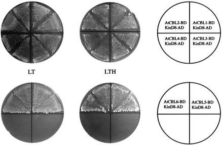
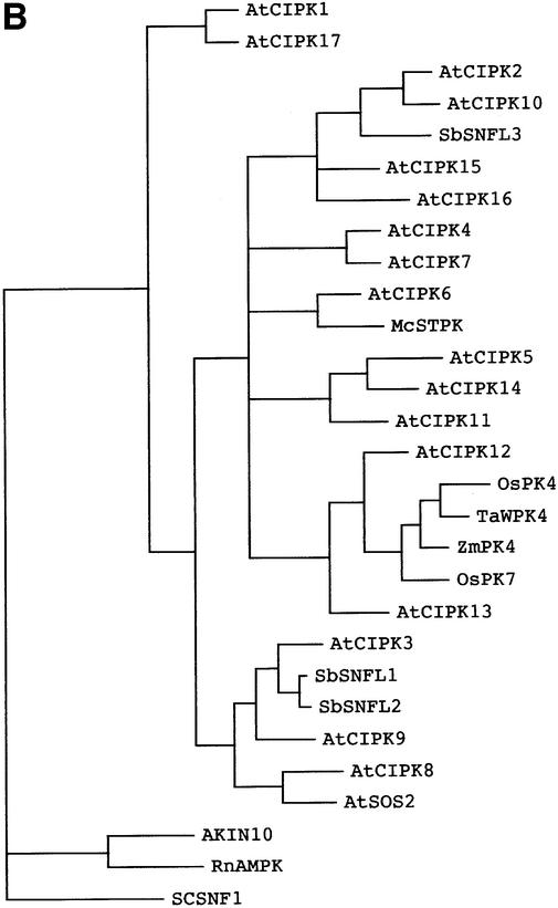
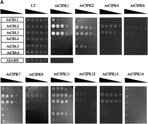
The Arabidopsis calcineurin B-like calcium sensor proteins (AtCBLs) interact with a group of serine-threonine protein kinases (AtCIPKs) in a calcium-dependent manner. Here we identify a 24 amino acid domain (NAF domain) unique to these kinases as being required and sufficient for interaction with all known AtCBLs. Mutation of conserved residues either abolished or significantly diminished the affinity of AtCIPK1 for AtCBL2. Comprehensive two-hybrid screens with various AtCBLs identified 15 CIPKs as potential targets of CBL proteins. Database analyses revealed additional kinases from Arabidopsis and other plant species harbouring the NAF interaction module. Several of these kinases have been implicated in various signalling pathways mediating responses to stress, hormones and environmental cues. Full-length CIPKs show preferential interaction with distinct CBLs in yeast and in vitro assays. Our findings suggest differential interaction affinity as one of the mechanisms generating the temporal and spatial specificity of calcium signals within plant cells and that different combinations of CBL-CIPK proteins contribute to the complex network that connects various extracellular signals to defined cellular responses.


Figures

None
Fig. 1. A 24 amino acid motif in AtCIPK1 is sufficient for interaction with AtCBL2. (A) The complete coding region (Kin) and different deletion constructs of AtCIPK1 (KinD1–9) were cloned into the pGAD.GH vector and introduced into yeast reporter strains containing the pGBD.AtCBL2 plasmid. Yeast growth (+ or –) was monitored on selective media (SC –Leu, –Trp, –His; supplemented with 25 mM 3-AT) and β-galactosidase activity was estimated as described in Materials and methods. The amino acid positions covered by each construct are indicated in the bars. The length of each peptide is depicted separately in parentheses. (B) Representative two-hybrid assay with the KinD8 construct. The arrangement of the yeast strains containing the different plasmids is indicated in the circle on the right. AD and BD refer to the Gal4 activation domain and binding domain plasmids, respectively. (C) In vitro interaction of AtCBL2 with a set of representative AtCIPK1 polypeptides. Left panel: autoradiograph of in vitro transcribed/translated Kin polypeptides. The indicated polypeptides were produced in reticulocyte lysate in the presence of [35S]methionine. A 0.5–2.0 µl aliquot of the translation reactions was separated by SDS–PAGE to analyse the in vitro expression of the polypeptides by autoradiography. Right panel: 35S-labelled Kin polypeptides (see left panel) and recombinant AtCBL2-His6 protein were co-incubated for 2 h on ice (+ AtCBL2). Co-purification was carried out on Ni-NTA–Sepharose and bound protein complexes were eluted with an EDTA-containing buffer. As a control, the assay was also performed without addition of recombinant protein (– AtCBL2). Eluted samples were separated on two separate SDS–polyacrylamide gels. One gel was autoradiographed (upper part) and the other was analysed by western blotting using an antiserum produced against AtCBL1 to verify AtCBL2-His6 purification (lower part). Molecular weight standards in kilodaltons are indicated on the right.
None
Fig. 2. Effect of mutations in the NAF domain of AtCIPK1 on binding to AtCBL2. (A) Mutational analysis of the NAF domain. The wild-type sequence of the NAF domain of AtCIPK1 is presented in the uppermost line (KinD8) with amino acid positions indicated on top. Amino acid residues substituted by mutagenesis are dark shaded. The conserved NAF motif is marked by a light grey background. The β-galactosidase activity was measured as described in Materials and methods. The measured activity is indicated in the graph on the right. (B) The yeast strain SMY3 harbouring the pGBD.AtCBL2 plasmid was transformed with vectors harbouring either the 24 residues of the wild-type NAF domain or the indicated mutated versions fused to the Gal4 activation domain. As controls, yeast cells were transformed with either vectors pGBD.BS and pGAD.GH or a combination of these vectors with the corresponding vectors expressing AtCBL2 or the NAF domain of AtCIPK1. The yeast cells were grown for 3 days at 30°C on SC medium lacking Leu and Trp (LT) or SC medium without Leu, Trp and His supplemented with 25 mM 3-AT. The array of the yeasts containing the different plasmids is indicated in the scheme on the right. (C) In vitro interaction of AtCBL2 with a set of representative KinD8 polypeptides. A 5 µg aliquot of recombinant AtCBL2-His6 per lane (+ AtCBL2) or control samples without recombinant protein (– AtCBL2) were run on two separate SDS–polyacrylamide gels and transferred to PVDF membranes. The first membrane was cut into three pieces (two lanes each) and each piece was incubated for 1 h with the indicated in vitro transcribed/translated, 35S-labelled KinD8 polypeptide. After washing, the membranes were analysed for bound kinase polypeptides by autoradiography (left panel). The second membrane was probed with an antiserum produced against AtCBL1 to confirm equal loading in all lanes (right panel). The arrows show the position of AtCBL2-His6. Molecular weight standards in kilodaltons are indicated on the right.
None
Fig. 3. The NAF domain mediates interaction with all known Arabidopsis CBL proteins. The yeast strain SMY3 containing the pGAD.KinD8 construct was transformed with pGBD.BS plasmids expressing the six different CBL proteins. The presence of both plasmids was monitored by growth on synthetic complete media lacking Leu and Trp (LT). Protein interaction was assayed on media lacking Leu, Trp and His, and supplemented with 25 mM 3-AT (LTH). The arrangement of the different yeast strains on the plates is depicted in the schemes on the right.
None
Fig. 4. The NAF domain represents a conserved protein–protein interaction module. (A) Alignment of the 24 amino acid polypeptide NAF domain of AtCIPK1, AtCIPK2, AtCIPK3 and AtCIPK4 (DDBJ/EMBL/GenBank accession Nos AF302112, AF286050, AF286051 and AY007221) corresponding to the protein–protein interaction domain identified in the interaction assays. Residues on a black background indicate conserved amino acids. (B) Amino acid alignment of the NAF domain of AtCIPK1 with the corresponding domains of WPK4 from wheat (TaWPK4), PK 4 and 7 from rice (OsPK4 and OsPK7), sorghum SNFL1–3 (SbSNFL1–3), PK4 from corn (ZmPK4) and STPK from ice plant (McSTPK). (DDBJ/EMBL/GenBank accession Nos for the complete proteins are: BAA34675.1, BAA83688.1, BAA83689.1, T14735, T14736, T14822, AAF22219.1 and AAD31900.) Amino acid residues conserved in the compared sequences are shown on a black background.
None
Fig. 5. The NAF domain of heterologous kinases interacts with AtCBL2. The cDNA fragment encoding the NAF domain of sorghum SNFL3 kinase was cloned into the pGAD.GH vector. pGAD.SNFL3D8 and pGBD.AtCBL2 plasmids (and several plasmid combinations used as controls) were introduced into the yeast strain SMY3 and the presence of both plasmids was selected for by growth on media without Leu and Trp (LT). Protein interaction was monitored by growth on media lacking Leu, Trp and His, and supplemented with 25 mM 3-AT (LTH). The plasmid combinations are indicated in the circle on the right.
None
Fig. 6. Sequence comparison and phylogenetic analysis of CIPK proteins. (A) Amino acid alignment of CIPK proteins from Arabidopsis. The depicted proteins correspond to the following DDBJ/EMBL/GenBank accession Nos: AtCIPK1 (AF302112), AtCIPK2 (AF286050), AtCIPK3 (AF286051), AtCIPK4 (AY007221), AtCIPK5 (AF285105), AtCIPK6 (AF285106), AtCIPK7 (AF290192), AtCIPK8 (AF290193), AtCIPK9 (AF295664), AtCIPK10 (AF295665), AtCIPK11 (AF295666), AtCIPK12 (AF295667), AtCIPK13 (AF295668), AtCIPK14 (AF295669) and AtCIPK15 (AF302111). The alignment was generated using the CLUSTAL method with DNASTAR software. Amino acids conserved in the 15 compared sequences are shown on a black background. The numbers on the right indicate the amino acid position. Dashes mark gaps introduced to improve the alignment. (B) Phylogenetic analysis of plant kinases containing the NAF domain. The alignment is based on the 444 amino acid complete coding region of AtCIPK1. The phylogenetic tree shown is a bootstrap parsimony 50% consensus tree. Phylogenetic analysis was performed with the PAUP 4.0 program package (http:/www.Ims.si.edu/PAUP/). For DDBJ/EMBL/GenBank accession numbers of AtCIPKs, see above. The other plant protein kinases containing a NAF domain are described in the legend to Figure 4B. Yeast SNF kinase (SCSNF1, accession No. NP010765), rat AMPK-activated protein kinase (RnAMPK, Q09137) and Arabidopsis SNF1-related protein kinase 10 (AKIN10, Q38997) were included in the analysis as representative related kinases.
None
Fig. 7. Specific formation of CBL–CIPK complexes in yeast two-hybrid assays and in vitro. (A) Yeast SMY3 strains containing the indicated plasmid combinations were grown in SC medium without Leu and Trp to an OD600 of 2.0, and 10 µl aliquots of different dilutions (1, 10–1–10–4) were spotted onto selective and non-selective plates (non-selective medium, SC –Leu, –Trp; selective medium, SC –His, –Leu, –Trp, supplemented with 25 mM 3-AT). The combination of plasmids is indicated on the left (pGBD.AtCBL1–6) and at the top (different pGAD.AtCIPKs). Decreasing cell densities in the dilution series are illustrated by narrowing triangles. LT depicts a representative dilution series on non-selective plates. All experiments were carried out at least in duplicate. (B) In vitro interaction of AtCBL2 and AtCBL6 with AtCIPK1 (lanes 1), AtCIPK13 (lanes 2) and AtCIPK14 (lanes 3). Left panel: autoradiograph of in vitro transcribed/translated AtCIPKs. The indicated kinases were produced as described in Figure 1C. A 2.0 µl aliquot of each of the translation reactions was separated by SDS–PAGE followed by autoradiography. Right panel: the indicated 35S-labelled AtCIPKs and recombinant His6-tagged AtCBL2 or AtCBL6 were co-incubated for 2 h on ice. As a control, the assay was also performed without added CBL protein (– AtCBL). Co-purification of CBL–CIPK complexes and autoradiography of the samples were carried out as described in Figure 1C. Affinity-purified AtCBL proteins were detected by Ni-NTA conjugate recognizing the His6 tag. Molecular weight standards in kilodaltons are indicated on the right.

References

    1. Annen F. and Stockhaus,J. (1998) Characterisation of a Sorghum bicolor gene family encoding putative protein kinases with a high similarity to the yeast SNF1 protein kinase. Plant Mol. Biol., 36, 529–539. - PubMed
    1. Bootman M.D., Berridge,M.J. and Lipp,P. (1997) Cooking with calcium: the recipes for composing global signals from elementary events. Cell, 91, 367–373. - PubMed
    1. Cardenas M.E., Hemenway,C., Muir,R.S., Ye,R., Fiorentino,D. and Heitman,J. (1994) Immunophilins interact with calcineurin in the absence of exogenous immunosuppressive ligands. EMBO J., 13, 5944–5957. - PMC - PubMed
    1. Chen C.-K., Inglese,J., Lefkowitz,R.J. and Hurley,J.B. (1995) Ca2+-dependent interaction of recoverin with rhodopsin kinase. J. Biol. Chem., 270, 18060–18066. - PubMed
    1. Clapham D.E. (1995) Calcium signalling. Cell, 80, 259–268. - PubMed

Publication types

MeSH terms