. 2001 Mar 1;20(5):1064-73.
doi: 10.1093/emboj/20.5.1064.

Fission yeast Pom1p kinase activity is cell cycle regulated and essential for cellular symmetry during growth and division

Affiliations

Fission yeast Pom1p kinase activity is cell cycle regulated and essential for cellular symmetry during growth and division

J Bähler et al. EMBO J. .

Abstract

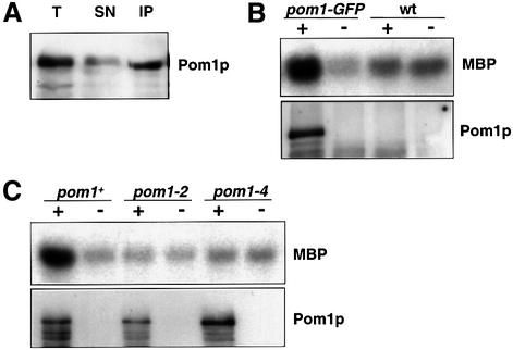
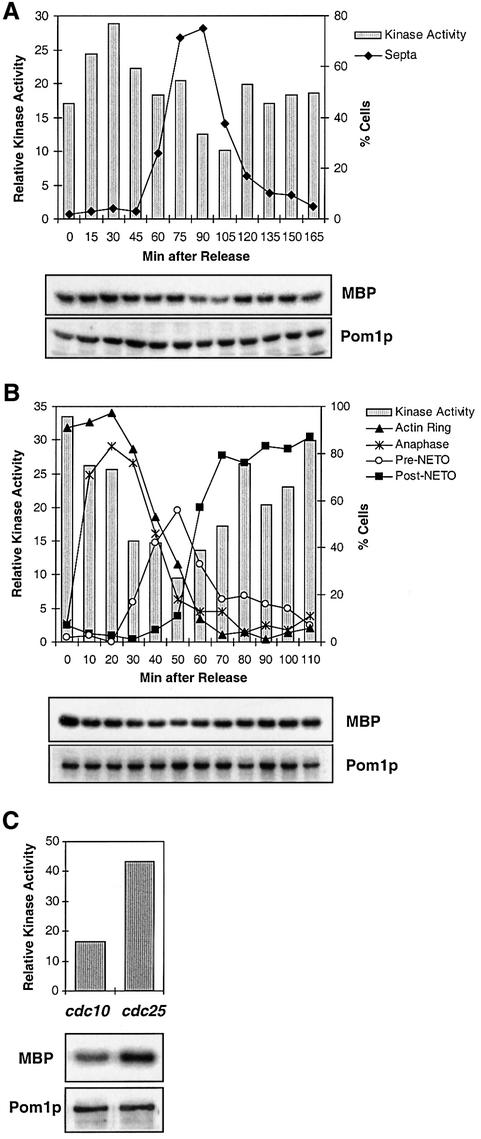
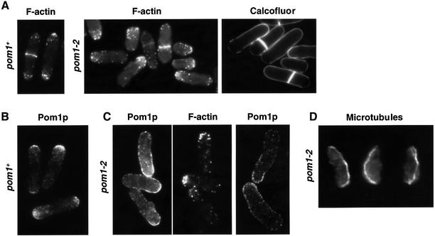
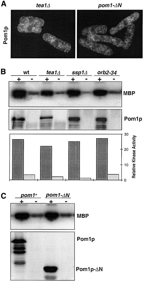
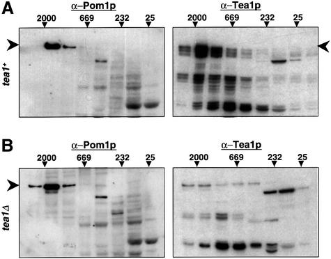
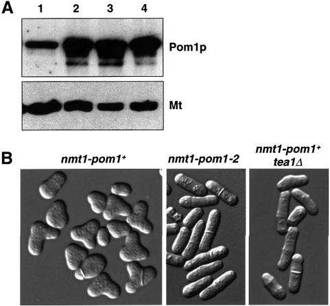
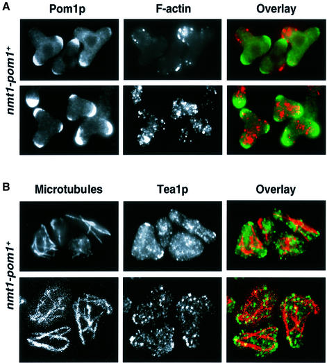
Schizosaccharomyces pombe cells grow from both ends during most of interphase and divide symmetrically into two daughter cells. The pom1 gene, encoding a member of the Dyrk family of protein kinases, has been identified through a mutant showing abnormal cellular morphogenesis. Here we show that Pom1p kinase activity is cell cycle regulated in correlation with the state of cellular symmetry: the activity is high during symmetrical growth and division, but lower when cells grow at just one end. Point mutations in the catalytic domain lead to asymmetry during both cell growth and division, whilst cells overexpressing Pom1p form additional growing ends. Manipulations of kinase activity indicate a negative role for Pom1p in microtubule growth at cell ends. Pom1p is present in a large protein complex and requires its non-catalytic domain to localize to the cell periphery and its kinase activity to localize to cell ends. These data establish that Pom1p kinase activity plays an important role in generating cellular symmetry and suggest that there may be related roles of homologous protein kinases ubiquitously present in all eukaryotes.


Figures

None
Fig. 1. In vitro protein kinase activity of Pom1p. (A) Immuno precipitation of Pom1p. Protein from strain JB115 (pom1-GFP) was extracted and precipitated using polyclonal α-GFP antibodies. The western blot shows aliquots of total extract (T), supernatant (SN) and precipitate (IP) loaded in equal quantities and probed with monoclonal α-GFP antibodies. Pom1p–GFP runs at ∼150 kDa. (B) Protein kinase assay of Pom1p. Protein extracts from strain JB115 (pom1-GFP) and the untagged 972 wild-type strain (wt) were precipitated in the presence (+) or absence (–) of α-GFP antibodies. The autoradiograph shows the Pom1p-associated phosphorylation of MBP, and western blot of the same samples probed with α-GFP antibodies to detect tagged Pom1p. (C) Absence of kinase activity in pom1 kinase mutants. Protein extracts of strains JB115 (pom1-GFP), and two mutant strains JB310 (pom1-2-GFP) and JB312 (pom1-4-GFP), which have point mutations in the ATP binding site and the activation loop, respectively, of the pom1 kinase domain, were precipitated in the presence (+) and absence (–) of α-GFP antibodies. Autoradiograph and western blot as in (B).
None
Fig. 2. Cell cycle regulation of Pom1p protein kinase activity. (A) The temperature-sensitive strain JB318 (pom1-GFP cdc25-22) was grown in EMM2 medium to OD595 0.15, shifted to the restrictive temperature (36°C) for 4 h and then released to the permissive temperature (25°C). Aliquots of cells were harvested immediately before and every 15 min after the shift to 25°C. At each time point, the percentage of septated cells was determined by Calcofluor staining. Protein extracts from each time point were precipitated with α-GFP antibodies and analysed for Pom1p kinase activity (MBP) and the presence of Pom1p. The kinase activities were quantified with a PhosphoImager and normalized for the amount of Pom1p present. (B) The cold-sensitive strain JB319 (pom1-GFP nda3-KM311) was grown in YE medium to OD595 0.3, shifted to the restrictive temperature (20°C) for 6 h and then released to the permissive temperature (32°C). Aliquots of cells were harvested immediately before and every 10 min after the shift to 32°C. For each time point, cell cycle stages were determined by DAPI staining to check for anaphase (presence of two nuclei) and by phalloidin staining to check for cytokinesis, pre- and post-NETO cells (presence of F-actin ring, unipolar and bipolar actin distribution, respectively). Pom1p kinase activities (MBP) and the presence of Pom1p were analysed at each time point as in (A). (C) The temperature-sensitive strains JB309 (pom1-GFP cdc10–129) and JB318 (pom1-GFP cdc25-22) were grown in EMM medium to OD595 0.2 and shifted to the restrictive temperature (36°C) for 3.5 h. Pom1p protein kinase activities in the arrested cells were determined as in (A). cdc10 and cdc25 cells arrest before and after the switch to bipolar growth, respectively (Mitchison and Nurse, 1985).
None
Fig. 3. Cellular functions of Pom1p protein kinase activity. (A) Defects in both symmetrical cell growth and cell division in a strain mutated in the pom1 kinase domain. Strain JB179 (pom1-2) was stained for F-actin with phalloidin and for cell wall growth with Calcofluor. Phalloidin-stained 972 wild-type cells (pom1+) are shown for comparison. Note that F-actin is concentrated at one end only and actin rings are placed off centre in the mutant cells. Consequently, cell wall growth is limited to one end and division septa are formed asymmetrically. (B) Localization of Pom1p to the cell ends. Immunofluorescence of strain JB115 (pom1+-GFP) using α-GFP antibodies. Note that Pom1p is concentrated at cell ends. (C) Pom1p fails to localize exclusively to cell ends in a strain mutated in the kinase domain. Immunofluorescence of strain JB310 (pom1-2-GFP) using α-GFP and α-actin antibodies. Note that the mutated Pom1p localizes in a wide area in one half of the cell opposite to the end where F-actin is concentrated. The confocal section on the right shows that Pom1p is located at the cell periphery without being restricted to the very cell end. (D) Microtubules bend around the cell end in the absence of Pom1p kinase activity. Immunofluorescence of strain JB179 (pom1-2) using α-tubulin antibody. Note that microtubules are curled around the broader new cell ends (bottom), extending to greater length than they would in wild-type cells.
None
Fig. 4. Pom1p kinase localization and regulation. (A) Pom1p requires its N-terminal half and Tea1p to localize to the cell ends. Immuno fluorescence of strain JB321 (pom1-GFP tea1Δ) using α-GFP antibodies (left) and of strain JB176 (3HA-pom1-ΔN) using α-HA antibodies (right). Note that Pom1p localizes in speckles throughout the cytoplasm in both strains. The dark round regions in the cell centre correspond to the nuclei. (B) Pom1p protein kinase activity is independent of three known genes that are required for bipolar growth. Protein extracts of strains JB115 (pom1-GFP, wt), JB321 (pom1-GFP tea1Δ), JB325 (pom1-GFP ssp1Δ) and JB324 (pom1-GFP orb2-34) were precipitated in the presence (+) or absence (–) of α-GFP antibodies. Autoradiograph showing the Pom1p-associated phosphorylation of MBP, and a western blot of the same samples probed with α-GFP antibodies to detect tagged Pom1p. The graph at the bottom shows the quantitation of kinase activities with a PhosphoImager in the presence (dark grey: normalized for the amount of Pom1p) and absence (light grey) of α-GFP antibodies. (C) Pom1p protein kinase activity is independent of its N-terminal half. Protein extracts of strains JB175 (3HA-pom1+) and JB176 (3HA-pom1-ΔN) were precipitated in the presence (+) or absence (–) of α-HA antibodies. Autoradiograph and western blot as in (B).
None
Fig. 5. Pom1p is present in a high molecular mass complex similar to but independent of Tea1p. (A) Protein extracts of strain JB115 (pom1-GFP) were fractionated by gel filtration. The fractions were analysed on western blots probed with α-GFP (to detect Pom1p) or α-Tea1p antibodies. The migration of molecular mass markers is shown on top (values in kilodaltons). Arrowheads indicate the positions of full-length Pom1p and Tea1p. Note that the Pom1p and Tea1p complexes migrate at a similar molecular mass. (B) Protein extracts of strain JB321 (pom1-GFP tea1Δ) were fractionated by gel filtration and analysed as in (A). The α-Tea1p antibody cross-reacts with various other proteins [compare to (A)].
None
Fig. 6. Overexpression of Pom1p protein kinase leads to cell branching. (A) Western blot probed with α-Pom1p antibody. Protein extracts of the following strains were loaded: (1) 972 (wild type); (2) JB151 (nmt1-pom1+); (3) JB314 (nmt1-pom1-2); (4) JB322 (nmt1-pom1+ tea1Δ). The same blot was probed with an α-tubulin antibody as a loading control (Mt). Strains with pom1 under the control of the nmt1 promoter were grown in the absence of thiamine for 19 h as described in (B). (B) Cells of strains JB151 (nmt1-pom1+), JB314 (nmt1-pom1-2) and JB322 (nmt1-pom1+ tea1Δ) were grown in EMM2 + thiamine, shifted to EMM2 without thiamine to induce the nmt1 promoter for 19 h at 32°C. Micrographs were taken using differential interference contrast microscopy. Note that cell branching depends both on Pom1p kinase activity and on Tea1p.
None
Fig. 7. Cytoskeletal aberrations in cells overexpressing Pom1p. (A) Immunofluorescence of strain JB151 (nmt1-pom1+) using α-Pom1p and α-actin antibodies. Note that at 16 h after induction of the nmt1 promoter (top row), Pom1p is strongly localized at the two normal cell ends, whereas F-actin is mainly concentrated in the branching cell end. At 19 h after induction of the nmt1 promoter (bottom row), Pom1p is localized at all three cell ends, and the F-actin localization has become depolarized. (B) Immunofluorescence of strain JB151 using α-tubulin and α-Tea1p antibodies. The nmt1 promoter was induced for 19 h. Immunofluorescence micrographs (top row) and confocal micrographs (bottom row) are shown. The microtubules are shorter and aberrantly organized, and Tea1p fails to become highly concentrated at the cell ends when Pom1p is overexpressed.

References

    1. Bähler J. and Peter,M. (2000) Cell polarity in yeast. In Drubin,D.G. (ed.), Cell Polarity. Oxford University Press, Oxford, UK, pp. 21–77.
    1. Bähler J. and Pringle,J.R. (1998) Pom1p, a fission yeast protein kinase that provides positional information for both polarized growth and cytokinesis. Genes Dev., 12, 1356–1370. - PMC - PubMed
    1. Bähler J., Steever,A.B., Wheatley,S., Wang,Y.-L., Pringle,J.R., Gould,K.L. and McCollum,D. (1998a) Role of Polo kinase and Mid1p in determining the site of cell division in fission yeast. J. Cell Biol., 143, 1603–1616. - PMC - PubMed
    1. Bähler J., Wu,J.-Q., Longtine,M.S., Shah,N.G., McKenzie,A.M.,III, Steever,A.B., Wach,A., Philippsen,P. and Pringle,J.R. (1998b) Heterologous modules for efficient and versatile PCR-based gene targeting in Schizosaccharomyces pombe. Yeast, 14, 943–951. - PubMed
    1. Becker W. and Joost,H.G. (1999) Structural and functional characteristics of Dyrk, a novel subfamily of protein kinases with dual specificity. Prog. Nucleic Acid Res. Mol. Biol., 62, 1–17. - PubMed

Publication types

MeSH terms

Substances