. 2001 Mar 1;20(5):1074-85.
doi: 10.1093/emboj/20.5.1074.

GSK-3 kinase Mck1 and calcineurin coordinately mediate Hsl1 down-regulation by Ca2+ in budding yeast

Affiliations

GSK-3 kinase Mck1 and calcineurin coordinately mediate Hsl1 down-regulation by Ca2+ in budding yeast

M Mizunuma et al. EMBO J. .

Abstract

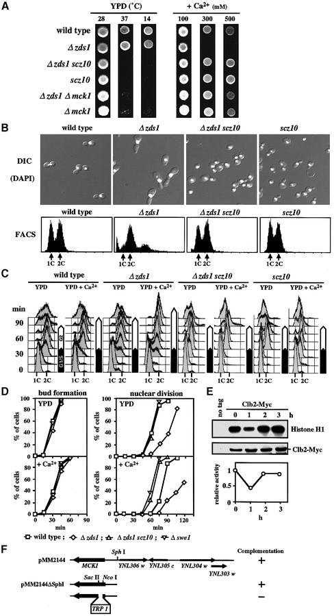
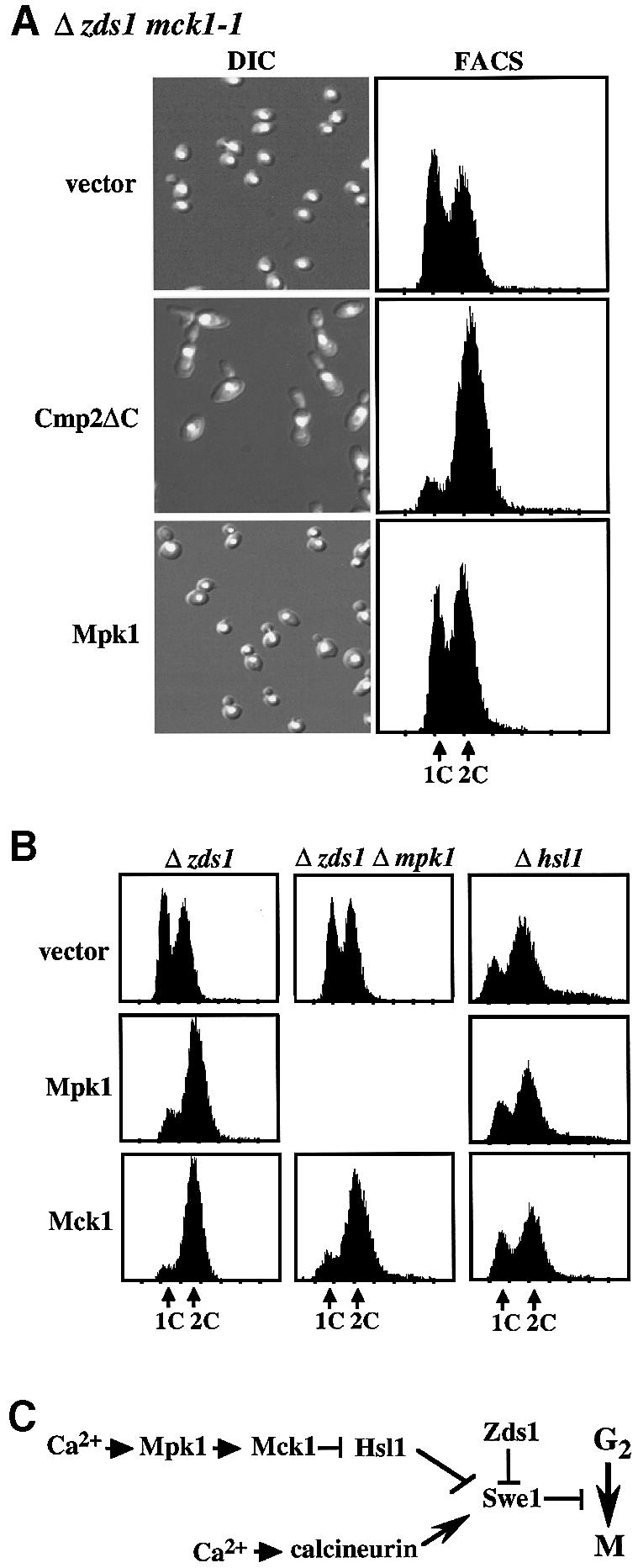
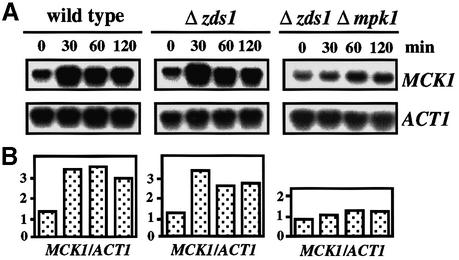
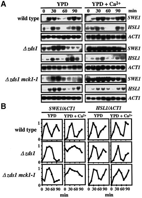
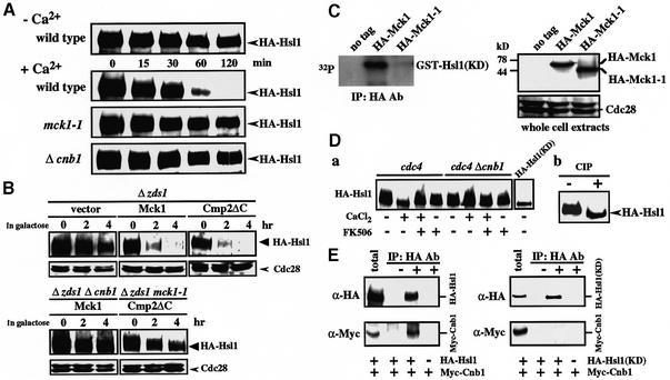
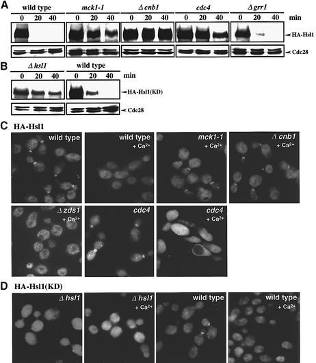
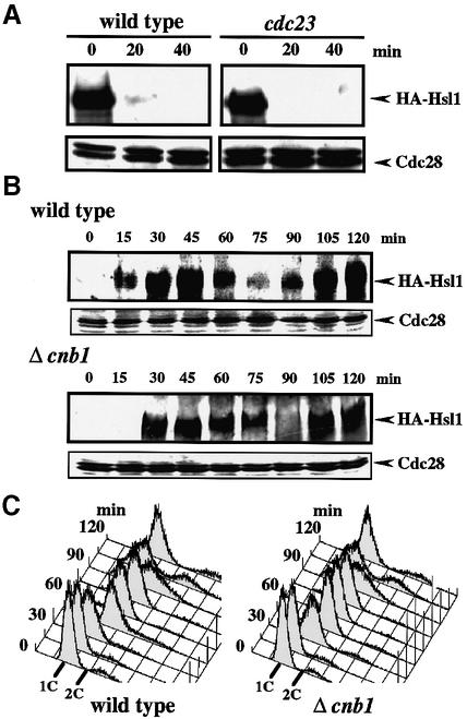
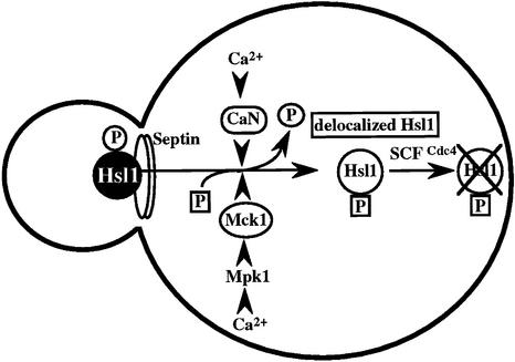
The Ca2+-activated pathways of Saccharomyces cerevisiae induce a delay in the onset of mitosis through the activation of Swe1, a negative regulatory kinase that inhibits the Cdc28-Clb complex. Calcineurin and Mpk1 activate Swe1 at the transcriptional and post-translational level, respectively, and both pathways are essential for the cell cycle delay. Our genetic screening identified the MCK1 gene, which encodes a glycogen synthetase kinase-3 family protein kinase, as a component of the Ca2+ signaling pathway. Genetic analyses indicated that Mck1 functions downstream of the Mpk1 pathway and down-regulates Hsl1, an inhibitory kinase of Swe1. In medium with a high concentration of Ca2+, Hsl1 was delocalized from the bud neck and destabilized in a manner dependent on both calcineurin and Mck1. Calcineurin was required for the dephosphorylation of autophosphorylated Hsl1. The E3 ubiquitin ligase complex SCF(Cdc4), but not the anaphase-promoting complex (APC), was essential for Hsl1 destabilization. The Ca2+-activated pathway may play a role in the rapid inactivation of Hsl1 at the cell cycle stage(s) when APC activity is low.


Figures

None
Fig. 1. The scz10 mutation suppresses various phenotypes of the Δzds1 strain. (A) Effect of scz10 (mck1-1) on the growth of the Δzds1 mutant strain on solid medium. Wild-type (DHT22-1b), Δzds1 (YAT1), Δzds1 scz10 (YMM2144), Δzds1Δmck1 (YMM69), scz10 (YMM22) or Δmck1 (YMM68) cells were spotted on YPD plates and grown at 28°C (2 days), 37°C (2 days) or 14°C (4 days), or on YPD plates supplemented with the indicated concentration of CaCl2 and incubated at 28°C for 2 days. (B) Cell morphology (DIC), DAPI fluorescence image and flow cytometry analysis of PI-stained cells (FACS: 1C, one DNA copy; or 2C, two DNA copies) of various strains after 6 h of incubation with 100 mM CaCl2 at 28°C. (C) DNA content during cell cycle progression in various strains. Early log phase growing cells (OD600 of 0.2–0.3) of DHT22-1b, YAT1, YMM2144 or YMM22 strains were synchronized with α-factor in G1, and resuspended in YPD or YPD plus 50 mM CaCl2. Samples were taken 15 min after removing α-factor. (D) Quantification of the cumulative percentage of bud formation and nuclear division in the cell cultures prepared as in (C). For comparison, the data with Δswe1 (YMM5-8c) are shown. Open squares, DHT22-1b; open diamonds, YAT1; open triangles, YMM2144; inverted open triangles, YMM5-8c. (E) Inhibition of Clb2–Cdc28 activity by CaCl2 in vivo. CaCl2 was added to a final concentration of 100 mM to early log phase culture of wild-type cells expressing (TMY310) or not expressing (DHT22-1b) Clb2-Myc in YPD at 28°C, and incubated further. The cell extracts prepared were immunoprecipitated with anti-Myc and assayed for histone H1 kinase acitivity. In parallel, cell extracts were immunoprecipitated with anti-Myc antibody, separated by SDS–PAGE and detected with Clb2-Myc. Activity was normalized relative to the Clb2 level of each lane. Activity was expressed relative to the activity at time zero (=1.0). (F) Map of the insert ORFs in plasmid pMM2144, which fully complemented the scz10 mutation. Deletion analysis was carried out to localize the suppressor activity (indicated by the + sign).
None
Fig. 2. Mck1 functions downstream of the Mpk1 pathway to induce G2 delay through the down-regulation of Hsl1. (A) The effect of overexpression of Cmp2ΔC (constitutively active form of the Cmp2 calcineurin catalytic subunit) or Mpk1 in the Δzds1 mck1-1 strain was examined. The Δzds1 mck1-1 (YMM2144) strain was transformed with the GAL-regulated plasmids pGAL-Cmp2ΔC (pYES2::Cmp2ΔC), pGAL-Mpk1 (pNV7::Mpk1) or vector alone (pYES2). The transformants were grown in liquid synthetic complete medium (SD minus Ura) at 28°C until early log growth phase. Cells were collected by centrifugation, washed, and suspended in SG (galactose medium, GAL promoter on) and the cells were then incubated at 28°C for 10 h. Cell morphology (DIC), FACS and the DNA content of PI-stained cells were analyzed by flow cytometry analysis. (B) The effect of overexpression of Mpk1 or Mck1 in Δzds1 (YAT1), Δzds1Δmpk1 (YMM3) or Δhsl1 (YMM52) strains was determined. These strains were transformed with pGAL-Mpk1 (pNV7::Mpk1), pGAL-Mck1 (pYES2::Mck1) or vector alone (pYES2). Experimental conditions were as described in (A). (C) Presumed pathway for the regulation of the Hsl1 kinase by Mck1 as suggested by the genetic analyses.
None
Fig. 3. Mpk1 up-regulates MCK1 transcription. CaCl2 was added to a final concentration of 100 mM to early log phase cultures of wild-type (DHT22-1b), Δzds1 (YAT1) or Δzds1 Δmpk1 (YMM3) cells in YPDat 28°C, and incubated further. RNA samples were extracted at the indicated time and northern blot analysis was performed. The intensity of MCK1 or ACT1 mRNA was measured using a BAS-1000 bio imaging analyzer and the MCK1 level was normalized to the ACT1 mRNA level.
None
Fig. 4. Mck1 is not involved in the transcriptional regulation of HSL1. (A) The oscillation of SWE1 or HSL1 mRNA levels during cell cycle progression in various strains that were synchronized with α-factor was determined by northern blotting, using the SWE1, HSL1 and ACT1 probes. Experimental conditions were as described in the legend to Figure 1C. RNA blot hybridization with the SWE1, HSL1 and ACT1 probes. (B) Changes in the relative intensity of SWE1 or HSL1 mRNA. The amount of SWE1 or HSL1 mRNA in (A) was normalized to a constant level of ACT1 mRNA. In each case, the individual maximum value was referred to as 1. Time 0 represents the point of release from G1 phase arrest.
None
Fig. 5. Mck1 and calcineurin cooperatively regulate Hsl1 abundance. (A) Comparison of Hsl1 abundance in various strains. Wild-type (YMM53), mck1-1 (YMM71) or Δcnb1 (YMM72), in which the genomic copy of HSL1 encodes HA-Hsl1, were grown in liquid YPD at 28°C until early log phase. CaCl2 was added to the cell cultures at the indicated times to a final concentration of 100 mM, samples were taken and western blot analysis was performed. (B) Dependence of Hsl1 destabilization on the functions of the calcineurin and Mpk1–Mck1 pathways. The strain YMM54, in which the genomic copy of HSL1 was replaced with HA-Hsl1, was transformed with the plasmids pGAL-Cmp2ΔC (pYES2::Cmp2ΔC), pGAL-Mck1 (pYES2::Mck1) or vector alone (pYES2). The transformants were grown in liquid YPR medium containing raffinose at 28°C until early log phase, and then galactose was added. Samples were taken at the indicated times and western blot analysis was performed by probing with anti-HA and anti-Cdc28 (control) antibodies. (C) Phosphorylation of Hsl1 by Mck1 in vitro. Cell extracts prepared from Δmck1 (YMM68) cells expressing HA-Mck1, HA-Mck1-1 or vector alone were immunoprecipitated with anti-HA. Kinase assay was performed by incubating the pellet with [γ-32P]ATP and recombinant GST–Hsl1(KD). Protein samples were subjected to electrophoresis and exposed for autoradiography. Western blotting was performed to detect HA-Mck1 and HA-Mck1-1, as well as Cdc28 as loading control. (D) (a) Mobility shift of HA-Hsl1 in the cells with activated Ca2+ signaling conditions in a calcineurin-dependent manner. Strain cdc4 (YMM73) or cdc4 Δcnb1 (YMM74), in which the genomic copy of HSL1 encodes HA-Hsl1, was incubated at 37°C for 2 h to inactivate cdc4. Incubation was carried out for 1 h in the medium with (+) or without (–) CaCl2 (100 mM) or FK506 (1 µg/ml) as indicated. HA-Hsl1 was detected by immunoblotting using anti-HA monoclonal antibody. (b) Phosphatase (CIP) treatment of HA-Hsl1 in the sample prepared similarly to the sample (+CaCl2, +FK506 of cdc4 cells) in (a). (E) Physical interaction of Hsl1 and calcineurin. Protein extracts prepared from the cells in which the genomic copy of HSL1 encodes HA-Hsl1 (YMM67), HA-Hsl1(KD) (YMM62) or none (DHT14) on a Δcnb1 background harboring plasmid-borne Myc-Cnb1 were immunoprecipitated with anti-HA antibody. The pellets were separated by SDS–PAGE and probed with both anti-HA and anti-Myc antibodies.
None
Fig. 6. Ca2+ induces SCFCdc4-mediated Hsl1 destabilization and Hsl1 delocalization from the bud neck in a manner dependent on both calcineurin and Mck1. (A) Comparison of Hsl1 abundance in various strains. Wild type (YMM53), mck1-1 (YMM71), Δcnb1 (YMM72), Δzds1 (YMM54), cdc4 (YMM73) or Δgrr1 (YMM76), each bearing a gene encoding HA-Hsl1, was grown in liquid YPD at 28°C until early log phase, and then shifted to 37°C for 2 h to inactivate Cdc4. CaCl2 was added to the cell cultures to a final concentration of 100 mM, samples were taken at intervals of 20 min and western blot analysis was performed. (B) Comparison of the Ca2+-induced destabilization of a kinase-negative Hsl1, HA-Hsl1(KD), in strains Δhsl1 (YMM60) or wild-type HSL1 (YMM66), each bearing a gene encoding HA-Hsl1(KD). Experimental conditions were as described in (A). (C) HA-Hsl1 localization in various strains. The cells were grown in liquid YPD at 28°C until early log phase, and then shifted to 37°C for 2 h to inactivate cdc4. CaCl2 was added to the cell cultures to a final concentration of 100 mM, and samples were then taken after 20 min of incubation. Indirect immunofluorescence microscopy was performed as described in Materials and methods. (D) Comparison of the localization of HA-Hsl1(KD) in Δhsl1 (YMM60) or HSL1 (YMM66) cells. CaCl2 was added to the cell cultures to a final concentration of 100 mM, and samples were then taken after 20 min of incubation. Experimental conditions were the same as in (C).
None
Fig. 7. The Ca2+-regulated Hsl1 destabilization pathway is distinct from the APC pathway. (A) Effect of Ca2+ on Hsl1 abundance in an APC mutant. Strains YMM53 or YMM65 (cdc23) with integrated HA-Hsl1 were grown in liquid YPD at 28°C until early log phase, and then shifted to 37°C for 2 h to inactivate cdc23. CaCl2 was added to the cell cultures to a final concentration of 100 mM, samples were taken at 20 min intervals and western blot analysis was performed. (B) The fluctuation of the Hsl1 abundance during the cell cycle in a calcineurin-deficient mutant. Strain YMM53 or YMM72 (Δcnb1), in which the genomic copy of HSL1 encodes an HA-tagged form of Hsl1, was grown in YPD to early log phase, and cells were synchronized as described in Figure 1C. Samples were taken at 15 min intervals after release in YPD, and whole-cell extracts were prepared. Immunoblotting was performed with antibodies against HA-Hsl1 and Cdc28 as loading control. (C) DNA content of YMM53 or YMM72 cells in a synchronous culture after release from an α-factor-induced G1 arrest.
None
Fig. 8. Model for the role of the Ca2+ signaling pathways of budding yeast in regulating the G2 phase delay through the delocalization and destabilization of Hsl1 from the bud neck. CaN, calcineurin.

Similar articles

Cited by

References

    1. Aberle H., Bauer,A., Stappert,J., Kispert,A. and Kemler,R. (1997) β-catenin is a target for the ubiquitin–proteasome pathway. EMBO J., 16, 3797–3804. - PMC - PubMed
    1. Amon A. (1997) Regulation of B-type cyclin proteolysis by Cdc28-associated kinase in budding yeast. EMBO J., 16, 2693–2702. - PMC - PubMed
    1. Amon A., Irniger,S. and Nasmyth,K. (1994) Closing the cell cycle circle in yeast: G2 cyclin proteolysis initiated at mitosis persists until the activation of G1 cyclins in the next cycle.Cell, 77, 1037–1050. - PubMed
    1. Andoh T., Hirata,Y. and Kikuchi,A. (2000) Yeast glycogen synthase kinase 3 is involved in protein degradation in cooperation with Bul1, Bul2 and Rsp5. Mol. Cell. Biol., 20, 6712–6720. - PMC - PubMed
    1. Barral Y., Parra,M., Bidlingmaier,S. and Snyder,M. (1999) Nim1-related kinases coordinate cell cycle progression with the organization of the peripheral cytoskeleton in yeast. Genes Dev., 13, 176–187. - PMC - PubMed

Publication types

MeSH terms

Substances