. 2001 Mar 1;20(5):1114-22.
doi: 10.1093/emboj/20.5.1114.

The transcription factors MTF-1 and USF1 cooperate to regulate mouse metallothionein-I expression in response to the essential metal zinc in visceral endoderm cells during early development

Affiliations

The transcription factors MTF-1 and USF1 cooperate to regulate mouse metallothionein-I expression in response to the essential metal zinc in visceral endoderm cells during early development

G K Andrews et al. EMBO J. .

Abstract

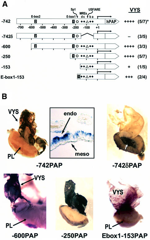
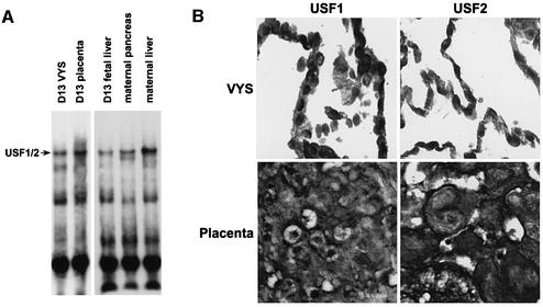
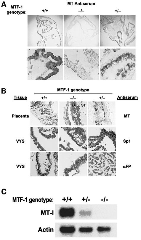
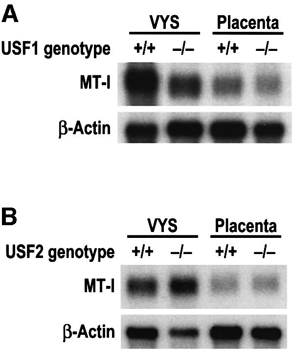
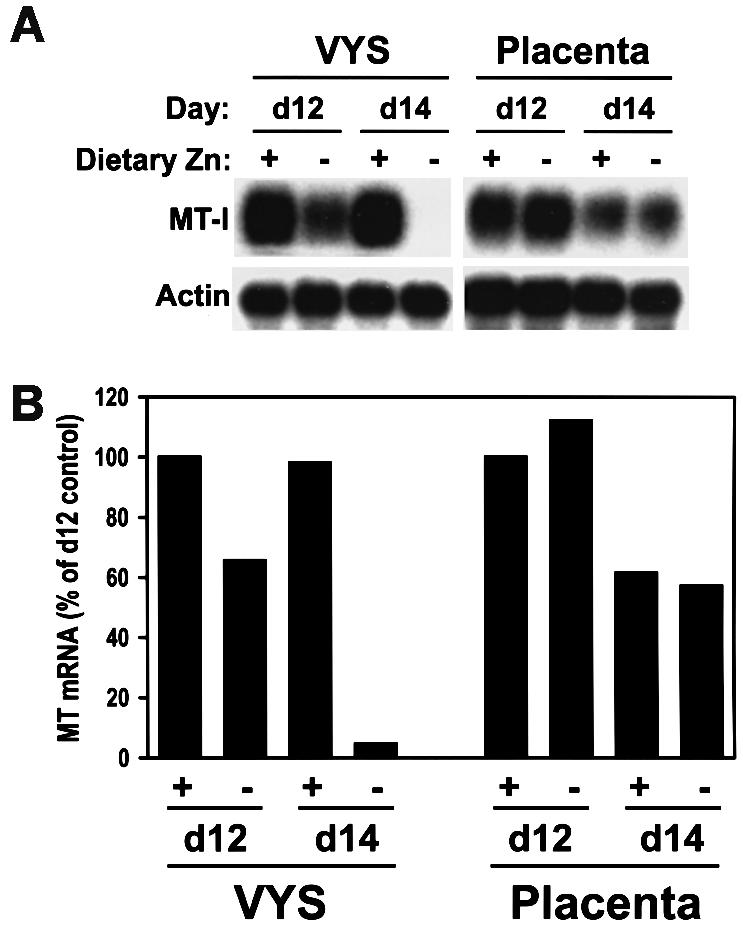
During early development of the mouse embryo, expression of the metallothionein-I (MT-I) gene is heightened specifically in the endoderm cells of the visceral yolk sac. The mechanisms of regulation of this cell-specific pattern of expression of metallothionein-I are unknown. However, it has recently been shown that MTF-1, functioning as a metalloregulatory transcription factor, activates metallothionein genes in response to the essential metal zinc. In contrast with the metallothionein genes, MTF-1 is essential for development; null mutant embryos die due to liver degeneration. We report here that MTF-1 is absolutely essential for upregulation of MT-I gene expression in visceral endoderm cells and that optimal expression also involves interactions of the basic helix-loop-helix upstream stimulatory factor-1 (USF1) with an E-box1-containing sequence at -223 bp in the MT-I promoter. Expression of MT-I in visceral endoderm cells was dependent on maternal dietary zinc. Thus, the essential metal, zinc, apparently provides the signaling ligand that activates cell-specific MT-I expression in visceral endoderm cells.


Figures

None
Fig. 1. Elements in the proximal promoter of mouse MT-I cooperate to direct high level hPAP reporter expression in the visceral yolk sac. (A) Diagrammatic representation of the mouse MT-I promoter–hPAP fusion constructs. The MT-I promoter provided the transcription start point (+1, straight arrow) and +66 bp of untranslated region, in addition to the base pairs of upstream promoter as indicated (Dalton et al., 1994). hPAP provided the translation start codon and complete coding sequence, and contains several introns (Knoll et al., 1988). A second polyadenylation signal is placed downstream of hPAP (Lin and Culp, 1991). Elements in the –742 bp promoter are as follows: E-box2 and E-box1, CAnnTG (Liang et al., 1996; Li et al., 1998); Sp1 GC-box (Briggs et al., 1986); MREs a–d, metal response elements (Stuart et al., 1984); USF/ARE, USF-binding site (not an E-box) overlapping an antioxidant response element (Dalton et al., 1994); TATA, TATA box. The –742δ promoter has a 108 bp region (bases –43 to –150 inclusive) deleted. The E-box1:–153 promoter has a single, forward-oriented copy of E-box1 inserted just upstream of the –153 bp MT-I promoter. hPAP, human placental alkaline phosphatase reporter; VYS, visceral yolk sac. The pattern of staining in the visceral yolk sac was assessed qualitatively [see (B)]: ++++, staining was always very dark and throughout the yolk sac endoderm; –, no cells stained in the visceral yolk sac in any transgenic embryos; +++, staining was dark in most cells; +, staining was weak and mosaic in the visceral yolk sac and detected in only one embryo. *Number of F0 transgenic conceptuses that expressed the hPAP gene/total number of transgenic embryos detected by PCR. (B) Histochemical detection of hPAP in F0 transgenic embryos. Promoter constructs are described above. VYS, visceral yolk sac; PL, placenta. Whole mounts of placentae were cross-sectioned before fixation and staining. The insert by the –742PAP whole mount is a photomicrograph of a thin section of the visceral yolk sac that shows hPAP expression in visceral endoderm (‘endo’) cells, but not in mesodermal cells (‘meso’).
None
Fig. 2. Visceral endoderm cell nuclei contain USF1 and USF2, which can bind in vitro to E-box1. (A) Nuclear proteins were prepared from day 13 visceral yolk sac, placenta and fetal liver, as well as from the maternal liver and pancreas. Nuclear proteins were analyzed by EMSA (Dalton et al., 1996c, 1997; Li et al., 1998) for E-box1-binding and for Sp1- and MTF-1-binding activities (data not shown). The E-box1-binding complex (USF1/2) contained USF1 and USF2 and was dependent on the E-box core-binding sequence (data not shown), as described (Li et al., 1998; Sirito et al., 1998). (B) Visceral yolk sacs and placentae were fixed and paraffin sections were analyzed for USF1 and USF2 proteins using immunohistochemistry. Nuclear staining was detected in the visceral yolk sac and placenta.
None
Fig. 3. Homozygous knockout of MTF-1 abolishes MT expression in the visceral endoderm. Heterozygous MTF-1 knockout mice were inbred and embryos were harvested at day 12 of gestation. The MTF-1 genotype of each embryo was determined by PCR. +/+, two normal MTF-1 alleles; +/–, heterozygous for MTF-1 knockout allele; –/–, homozygous for MTF-1 knockout alleles. The visceral yolk sacs and placentae were fixed and analyzed individually for MT, Sp1 or α-fetoprotein (αFP) using immunohistochemistry (A and B). D, deciduum basalis, ST, spongiotrophoblast layer. (A) Low- (100×) and high- (200×) magnification views of the immunohistochemical staining of MT in the visceral yolk sac. (C) Total RNA was extracted from the visceral yolk sac (four embryos) and analyzed by northern blot hybridization using MT-I and actin cRNA probes simultaneously on the same blot.
None
Fig. 4. Homozygous knockout of USF1, but not USF2, attenuates MT expression in the visceral endoderm. Heterozygous USF1 (A) or USF2 (B) knockout and control embryos and tissues were harvested at day 14 of gestation. +/+, embryo with two normal USF alleles; –/–, embryo homozygous for USF knockout alleles. Total RNA was extracted from the visceral yolk sac (VYS) and placenta (six per genotype) and analyzed by northern blot hybridization using MT-I and actin cRNA probes simultaneously on the same blot. Hybrids were quantified by radioanalytical analysis of the membranes. These RNA samples were northern blotted at least three separate times with similar results.
None
Fig. 5. Dietary zinc modulates MT-I expression in the visceral yolk sac. CD-1 mice were exposed to dietary zinc deficiency beginning on day 8 of pregnancy and the effects on MT-I mRNA levels in the visceral yolk sac and placenta were determined on days 12 and 14. Under these conditions, ∼20% of the embryos developed abnormally by day 14, but there was no increase in embryo mortality (Andrews and Geiser, 1999). Total RNA was extracted from visceral yolk sacs (VYS) and placentae (∼20 per sample) collected from mice fed a zinc-adequate (Zn-A) diet (+, containing 50 p.p.m. Zn2+) or a zinc-deficient (Zn-D) diet (−, containing 1 p.p.m. Zn2+). RNA was analyzed by northern blot hybridization using mouse MT-I and actin cRNA probes simultaneously. Hybrids were detected by autoradiography (A) and quantified by radioanalytic analysis of the membrane (B).

Similar articles

Cited by

References

    1. Andrews G.K. (2000) Regulation of metallothionein gene expression by oxidative stress and metal ions. Biochem. Pharmacol., 59, 95–104. - PubMed
    1. Andrews G.K. and Adamson,E.D. (1987) Butyrate selectively activates the metallothionein gene in teratocarcinoma cells and induces hypersensitivity to metal induction. Nucleic Acids Res., 15, 5461–5475. - PMC - PubMed
    1. Andrews G.K. and Geiser,J. (1999) Expression of metallothionein-I and -II genes provides a reproductive advantage during maternal dietary zinc deficiency. J. Nutr., 129, 1643–1648. - PubMed
    1. Andrews G.K., Adamson,E.D. and Gedamu,L. (1984) The ontogeny of expression of murine metallothionein: comparison with the α-fetoprotein gene. Dev. Biol., 103, 294–303. - PubMed
    1. Andrews G.K., Gallant,K.R. and Cherian,M.G. (1987a) Regulation of the ontogeny of rat liver metallothionein mRNA by zinc. Eur. J. Biochem., 166, 527–531. - PubMed

Publication types

MeSH terms